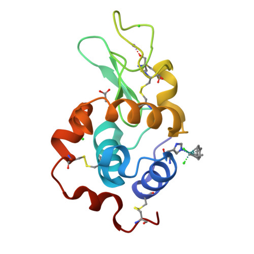
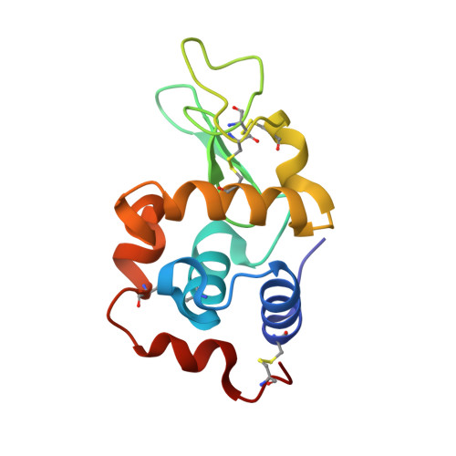
Half-sandwich arene ruthenium(II)-enzyme complex
McNae, I.W., Fishburne, K., Habtemariam, A., Hunter, T.M., Melchart, M., Wang, F., Walkinshaw, M.D., Sadler, P.J.(2004) Chem Commun (Camb) 16: 1786-1787
- PubMed: 15306883
- DOI: https://doi.org/10.1039/b408141b
- Primary Citation of Related Structures:
1T3P - PubMed Abstract:
The 1.6 [Angstrom] X-ray crystal structure of [(eta(6)-p-cymene)Ru(lysozyme)Cl(2)], the first of a half-sandwich complex of a protein, shows selective ruthenation of Nepsilon of the imidazole ring of His15.
Organizational Affiliation:
Institute of Cell and Molecular Biology, Michael Swann Building, University of Edinburgh, Mayfield Road, Edinburgh, UK.