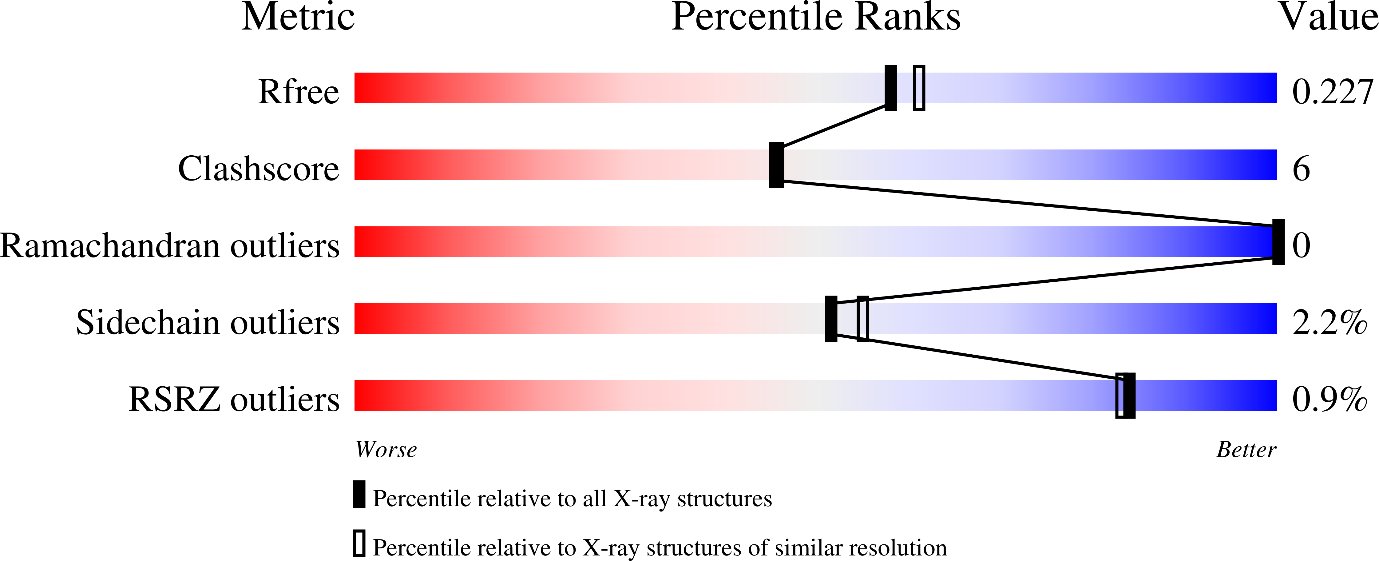
1QY7

The structure of the PII protein from the cyanobacteria Synechococcus sp. PCC 7942


Experimental Data Snapshot

  • Method: X-RAY DIFFRACTION
  • Resolution: 2.00 Å
  • R-Value Free: 0.227 
  • R-Value Work: 0.199 

Starting Model: experimental
View more details

wwPDB Validation   3D Report Full Report


This is version 1.4 of the entry. See complete history


Literature

The structures of the PII proteins from the cyanobacteria Synechococcus sp. PCC 7942 and Synechocystis sp. PCC 6803.

Xu, Y.Carr, P.D.Clancy, P.Garcia-Dominguez, M.Forchhammer, K.Florencio, F.Vasudevan, S.G.Tandeau de Marsac, N.Ollis, D.L.

(2003) Acta Crystallogr D Biol Crystallogr 59: 2183-2190

  • DOI: https://doi.org/10.1107/s0907444903019589
  • Primary Citation of Related Structures:  
    1QY7, 1UL3

  • PubMed Abstract: 

    The PII proteins from the cyanobacteria Synechococcus sp. PCC 7942 and Synechocystis sp. PCC 6803 have been crystallized and high-resolution structures have been obtained using X-ray crystallography. The core of these new structures is similar to that of the PII proteins from Escherichia coli, although the structures of the T- and C-loops differ. The T-loop of the Synechococcus protein is ordered, but appears to be stabilized by crystal contacts. The same loop in the Synechocystis protein is disordered. The C-terminus of the Synechocystis protein is stabilized by hydrogen bonding to the same region of a crystallographically related molecule. The same terminus in the Synechococcus protein is stabilized by coordination with a metal ion. These observations are consistent with the idea that both the T-loop and the C-terminus of PII proteins are flexible in solution and that this flexibility may be important for receptor recognition. Sequence comparisons are used to identify regions of the sequence unique to the cyanobacteria.


  • Organizational Affiliation

    Department of Biochemistry and Molecular Biology, James Cook University, Townsville, Queensland 4811, Australia.


Macromolecules
Find similar proteins by:  (by identity cutoff)  |  3D Structure
Entity ID: 1
MoleculeChains Sequence LengthOrganismDetailsImage
Nitrogen regulatory protein P-II
A, B, C
112Synechococcus elongatus PCC 7942 = FACHB-805Mutation(s): 1 
Gene Names: GLNB
UniProt
Find proteins for P0A3F4 (Synechococcus elongatus (strain ATCC 33912 / PCC 7942 / FACHB-805))
Explore P0A3F4 
Go to UniProtKB:  P0A3F4
Entity Groups  
Sequence Clusters30% Identity50% Identity70% Identity90% Identity95% Identity100% Identity
UniProt GroupP0A3F4
Sequence Annotations
Expand
  • Reference Sequence
Small Molecules
Ligands 2 Unique
IDChains Name / Formula / InChI Key2D Diagram3D Interactions
SO4
Query on SO4

Download Ideal Coordinates CCD File 
D [auth A]
E [auth A]
G [auth B]
H [auth B]
J [auth C]
D [auth A],
E [auth A],
G [auth B],
H [auth B],
J [auth C],
K [auth C]
SULFATE ION
O4 S
QAOWNCQODCNURD-UHFFFAOYSA-L
NI
Query on NI

Download Ideal Coordinates CCD File 
F [auth A],
I [auth B],
L [auth C]
NICKEL (II) ION
Ni
VEQPNABPJHWNSG-UHFFFAOYSA-N
Experimental Data & Validation

Experimental Data

  • Method: X-RAY DIFFRACTION
  • Resolution: 2.00 Å
  • R-Value Free: 0.227 
  • R-Value Work: 0.199 
  • Space Group: I 41 2 2
Unit Cell:
Length ( Å )Angle ( ˚ )
a = 109.7α = 90
b = 109.7β = 90
c = 109.68γ = 90
Software Package:
Software NamePurpose
DENZOdata reduction
SCALEPACKdata scaling
MOLREPphasing
CNSrefinement

Structure Validation

View Full Validation Report


Entry History 

Deposition Data

Revision History  (Full details and data files)

  • Version 1.0: 2003-09-23
    Type: Initial release
  • Version 1.1: 2008-04-29
    Changes: Version format compliance
  • Version 1.2: 2011-07-13
    Changes: Source and taxonomy, Version format compliance
  • Version 1.3: 2021-11-10
    Changes: Database references, Derived calculations
  • Version 1.4: 2023-10-25
    Changes: Data collection, Refinement description