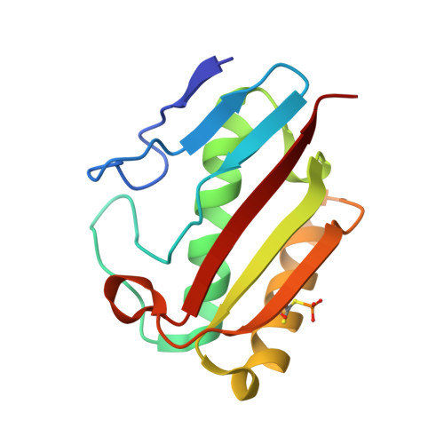
1QU9

1.2 A CRYSTAL STRUCTURE OF YJGF GENE PRODUCT FROM E. COLI


Experimental Data Snapshot

  • Method: X-RAY DIFFRACTION
  • Resolution: 1.20 Å
  • R-Value Work: 0.164 

wwPDB Validation   3D Report Full Report


This is version 1.4 of the entry. See complete history


Literature

A test case for structure-based functional assignment: the 1.2 A crystal structure of the yjgF gene product from Escherichia coli

Volz, K.

(1999) Protein Sci 8: 2428-2437

  • DOI: https://doi.org/10.1110/ps.8.11.2428
  • Primary Citation of Related Structures:  
    1QU9

  • PubMed Abstract: 

    The YER057c/YIL051c/YjgF protein family is a set of 24 full-length homologs, each approximately 130 residues in length, and each with no known function or relationship to proteins of known structure. To determine the function of this family, the structure of one member--the YjgF protein from Escherichia coli--was solved and refined at a resolution of 1.2 A. The YjgF molecule is a homotrimer with exact threefold symmetry. Its tertiary and quaternary structures are related to that of Bacillus subtilis chorismate mutase, although their active sites are completely different. The YjgF protein has an active site curiously similar to protein tyrosine phosphatases, including a covalently modified cysteine, but it is unlikely to be functionally related. The lessons learned from this attempt to deduce function from structure may be useful to future projects in structural genomics.


  • Organizational Affiliation

    Department of Microbiology and Immunology, University of Illinois at Chicago, 60612-7344, USA. [email protected]


Macromolecules
Find similar proteins by:  (by identity cutoff)  |  3D Structure
Entity ID: 1
MoleculeChains Sequence LengthOrganismDetailsImage
YJGF PROTEIN
A, B, C
128Escherichia coliMutation(s): 1 
EC: 3.5.99.10
UniProt
Find proteins for P0AF93 (Escherichia coli (strain K12))
Explore P0AF93 
Go to UniProtKB:  P0AF93
Entity Groups  
Sequence Clusters30% Identity50% Identity70% Identity90% Identity95% Identity100% Identity
UniProt GroupP0AF93
Sequence Annotations
Expand
  • Reference Sequence
Small Molecules
Modified Residues  1 Unique
IDChains TypeFormula2D DiagramParent
CSP
Query on CSP
A, B, C
L-PEPTIDE LINKINGC3 H8 N O5 P SCYS
Experimental Data & Validation

Experimental Data

  • Method: X-RAY DIFFRACTION
  • Resolution: 1.20 Å
  • R-Value Work: 0.164 
  • Space Group: P 2 2 2
Unit Cell:
Length ( Å )Angle ( ˚ )
a = 72.22α = 90
b = 72.22β = 90
c = 72.22γ = 90
Software Package:
Software NamePurpose
MLPHAREphasing
PROFFTrefinement
SMARTdata reduction
SAINTdata reduction
SMARTdata scaling
SAINTdata scaling

Structure Validation

View Full Validation Report


Entry History 

Deposition Data

  • Released Date: 1999-12-01 
  • Deposition Author(s): Volz, K.

Revision History  (Full details and data files)

  • Version 1.0: 1999-12-01
    Type: Initial release
  • Version 1.1: 2008-04-27
    Changes: Version format compliance
  • Version 1.2: 2011-07-13
    Changes: Version format compliance
  • Version 1.3: 2018-01-31
    Changes: Experimental preparation
  • Version 1.4: 2024-11-20
    Changes: Data collection, Database references, Derived calculations, Structure summary