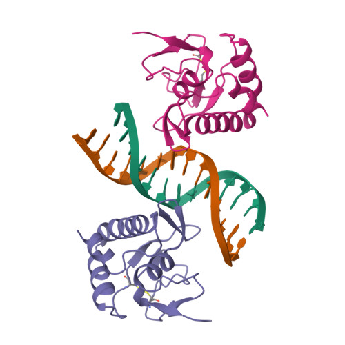
Crystal structure of a Smad MH1 domain bound to DNA: insights on DNA binding in TGF-beta signaling.
Shi, Y., Wang, Y.F., Jayaraman, L., Yang, H., Massague, J., Pavletich, N.P.(1998) Cell 94: 585-594
- PubMed: 9741623
- DOI: https://doi.org/10.1016/s0092-8674(00)81600-1
- Primary Citation of Related Structures:
1MHD - PubMed Abstract:
The Smad family of proteins, which are frequently targeted by tumorigenic mutations in cancer, mediate TGF-beta signaling from cell membrane to nucleus. The crystal structure of a Smad3 MH1 domain bound to an optimal DNA sequence determined at 2.8 A resolution reveals a novel DNA-binding motif. In the crystals, base-specific DNA recognition is provided exclusively by a conserved 11-residue beta hairpin that is embedded in the major groove of DNA. A surface loop region, to which tumorigenic mutations map, has been identified as a functional surface important for Smad activity. This structure establishes a framework for understanding how Smad proteins may act in concert with other transcription factors in the regulation of TGF-beta-responsive genes.
Organizational Affiliation:
Department of Molecular Biology, Princeton University, Lewis Thomas Laboratory, New Jersey 08544, USA. ygshi@princeton.edu