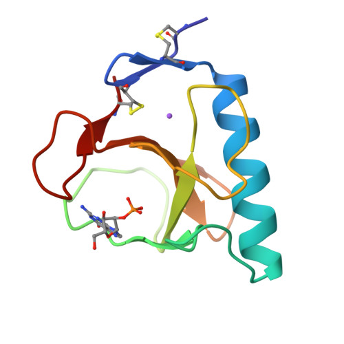
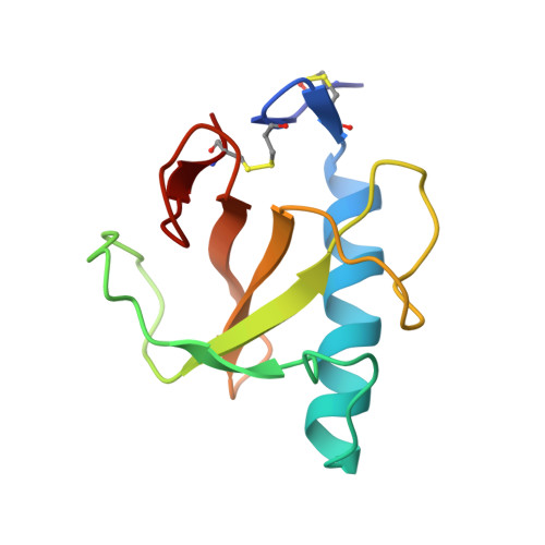
Crystallographic study of Glu58Ala RNase T1 x 2'-guanosine monophosphate at 1.9-A resolution.
Pletinckx, J., Steyaert, J., Zegers, I., Choe, H.W., Heinemann, U., Wyns, L.(1994) Biochemistry 33: 1654-1662
- PubMed: 7906540
- DOI: https://doi.org/10.1021/bi00173a006
- Primary Citation of Related Structures:
1LRA - PubMed Abstract:
Glu58 is known to participate in phosphodiester transesterification catalyzed by the enzyme RNase T1. For Glu58 RNase T1, an altered mechanism has been proposed in which His40 replaces Glu58 as the base catalyst [Steyaert, J., Hallenga, K., Wyns, L., & Stanssens, P. (1990) Biochemistry 29, 9064-9072]. Glu58Ala Rnase T1 has been cocrystallized with guanosine 2'-monophosphate (2'-GMP). The crystals are of space group P2(1), with one molecule per asymmetric unit (a = 32.44 A, b = 49.64 A, c = 26.09 A, beta = 99.17 degrees). The three-dimensional structure of the enzyme was determined to a nominal resolution of 1.9 A, yielding a crystallographic R factor of 0.178 for all X-ray data. Comparison of this structure with wild-type structures leads to the following conclusions. The minor changes apparent in the tertiary structure can be explained by either the mutation of Glu58 or by the change in the space group. In the active site, the extra space available through the mutation of Glu58 is occupied by the phosphate group (after a reorientation) and by a solvent molecule replacing a carboxylate oxygen of Glu58. This solvent molecule is a candidate for participation in the altered mechanism of this mutant enzyme. Following up on a study of conserved water sites in RNase T1 crystallized in space group P2(1)2(1)2(1) [Malin, R., Zielenkiewicz, P., & Saenger, W. (1991) J. Mol. Biol. 266, 4848-4852], we investigated the hydration structure for four different packing modes of RNase T1.(ABSTRACT TRUNCATED AT 250 WORDS)
Organizational Affiliation:
Instituut voor Moleculaire Biologie, Vrije Universiteit Brussel, Belgium.