1AJH | pdb_00001ajh

PHOTOPRODUCT OF CARBONMONOXY MYOGLOBIN AT 40 K


Experimental Data Snapshot

  • Method: X-RAY DIFFRACTION
  • Resolution: 1.69 Å
  • R-Value Free: 
    0.234 (Depositor) 
  • R-Value Work: 
    0.171 (Depositor), 0.170 (DCC) 
  • R-Value Observed: 
    0.171 (Depositor) 

Starting Model: experimental
View more details

wwPDB Validation   3D Report Full Report


Ligand Structure Quality Assessment 

Created with Raphaël 2.3.0Worse 01 BetterLigand structure goodness of fit to experimental dataBest fitted HEMClick on this verticalbar to view details

This is version 1.4 of the entry. See complete history


Literature

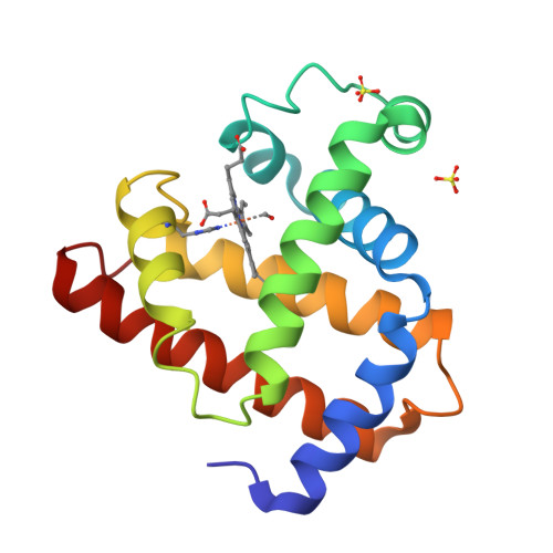
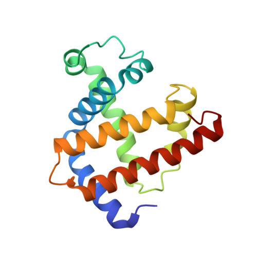
Photolysis-induced structural changes in single crystals of carbonmonoxy myoglobin at 40 K.

Teng, T.Y.Srajer, V.Moffat, K.

(1994) Nat Struct Biol 1: 701-705

  • DOI: https://doi.org/10.1038/nsb1094-701
  • Primary Citation of Related Structures:  
    1AJG, 1AJH

  • PubMed Abstract: 

    Myoglobin's reversible binding of oxygen is a model for studies of protein control of ligand binding and discrimination. Protein relaxation and geminate ligand rebinding subsequent to ligand photodissociation have been studied extensively by a variety of techniques. The ps to ns time scales for these processes are still much shorter than the ms time resolution of X-ray diffraction experiments, but it may be possible to trap these intermediates at low temperatures. We report here an X-ray diffraction investigation of structural changes induced by photolysis of carbonmonoxy myoglobin crystals at 40 K. Our results provide a structural basis for the interpretation of ambient and low temperature spectroscopic observations and molecular dynamics simulations of the ligand photodissociation and binding processes in haem proteins.


  • Organizational Affiliation

    Department of Biochemistry and Molecular Biology, University of Chicago, IL 60637, USA.


Macromolecules
Find similar proteins by:  (by identity cutoff)  |  3D Structure
Entity ID: 1
MoleculeChains Sequence LengthOrganismDetailsImage
MYOGLOBIN153Physeter macrocephalusMutation(s): 0 
EC: 1.11.1 (UniProt), 1.7 (UniProt)
UniProt
Find proteins for P02185 (Physeter macrocephalus)
Explore P02185 
Go to UniProtKB:  P02185
Entity Groups  
Sequence Clusters30% Identity50% Identity70% Identity90% Identity95% Identity100% Identity
UniProt GroupP02185
Sequence Annotations
Expand
  • Reference Sequence
Experimental Data & Validation

Experimental Data

  • Method: X-RAY DIFFRACTION
  • Resolution: 1.69 Å
  • R-Value Free:  0.234 (Depositor) 
  • R-Value Work:  0.171 (Depositor), 0.170 (DCC) 
  • R-Value Observed: 0.171 (Depositor) 
Space Group: P 1 21 1
Unit Cell:
Length ( Å )Angle ( ˚ )
a = 63.45α = 90
b = 30.44β = 105.7
c = 34.13γ = 90
Software Package:
Software NamePurpose
X-PLORmodel building
X-PLORrefinement
DENZOdata reduction
CCP4data scaling
X-PLORphasing

Structure Validation

View Full Validation Report


Ligand Structure Quality Assessment 

Created with Raphaël 2.3.0Worse 01 BetterLigand structure goodness of fit to experimental dataBest fitted HEMClick on this verticalbar to view details

Entry History 

Deposition Data

Revision History  (Full details and data files)

  • Version 1.0: 1997-11-12
    Type: Initial release
  • Version 1.1: 2008-03-24
    Changes: Version format compliance
  • Version 1.2: 2011-07-13
    Changes: Version format compliance
  • Version 1.3: 2023-08-02
    Changes: Database references, Derived calculations, Other, Refinement description
  • Version 1.4: 2024-05-22
    Changes: Data collection