7WUX | pdb_00007wux

Crystal structure of AziU3/U2 complexed with (5S,6S)-O7-sulfo DADH from Streptomyces sahachiroi


Experimental Data Snapshot

  • Method: X-RAY DIFFRACTION
  • Resolution: 1.80 Å
  • R-Value Free: 
    0.193 (Depositor), 0.200 (DCC) 
  • R-Value Work: 
    0.164 (Depositor), 0.170 (DCC) 

wwPDB Validation   3D Report Full Report


Ligand Structure Quality Assessment 

Created with Raphaël 2.3.0Worse 01 BetterLigand structure goodness of fit to experimental dataBest fitted 6OIClick on this verticalbar to view details

This is version 1.2 of the entry. See complete history


Literature

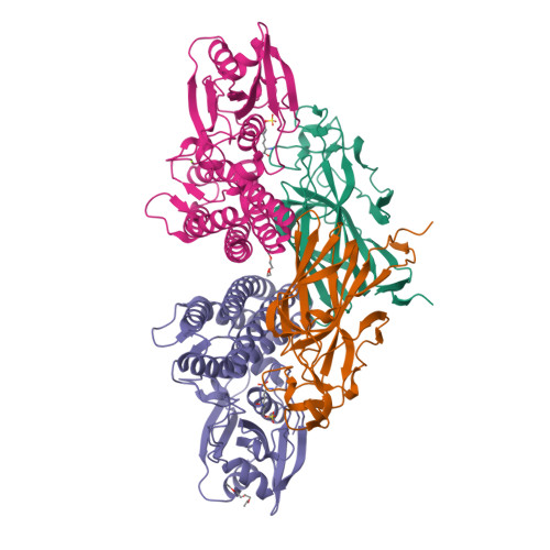
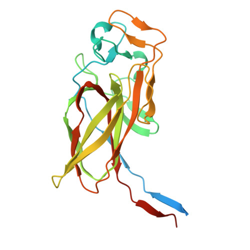
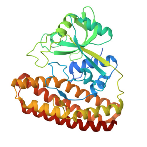
Molecular Basis for Enzymatic Aziridine Formation via Sulfate Elimination.

Kurosawa, S.Hasebe, F.Okamura, H.Yoshida, A.Matsuda, K.Sone, Y.Tomita, T.Shinada, T.Takikawa, H.Kuzuyama, T.Kosono, S.Nishiyama, M.

(2022) J Am Chem Soc 144: 16164-16170

  • DOI: https://doi.org/10.1021/jacs.2c07243
  • Primary Citation of Related Structures:  
    7WUW, 7WUX

  • PubMed Abstract: 

    Natural products containing an aziridine ring, such as mitomycin C and azinomycin B, exhibit antitumor activities by alkylating DNA via their aziridine rings; however, the biosynthetic mechanisms underlying the formation of these rings have not yet been elucidated. We herein investigated the biosynthesis of vazabitide A, the structure of which is similar to that of azinomycin B, and demonstrated that Vzb10/11, with no similarities to known enzymes, catalyzed the formation of the aziridine ring via sulfate elimination. To elucidate the detailed reaction mechanism, crystallization of Vzb10/11 and the homologous enzyme, AziU3/U2, in the biosynthesis of azinomycin B was attempted, and the structure of AziU3/U2, which had a new protein fold overall, was successfully determined. The structural analysis revealed that these enzymes adjusted the dihedral angle between the amino group and the adjacent sulfate group of the substrate to almost 180° and enhanced the nucleophilicity of the C6-amino group temporarily, facilitating the S N 2-like reaction to form the aziridine ring. The present study reports for the first time the molecular basis for aziridine ring formation.


  • Organizational Affiliation

    Graduate School of Agricultural and Life Sciences, The University of Tokyo, 1-1-1, Yayoi, Bunkyo-ku, Tokyo 113-8657, Japan.


Macromolecules
Find similar proteins by:  (by identity cutoff)  |  3D Structure
Entity ID: 1
MoleculeChains Sequence LengthOrganismDetailsImage
AziU2
A, B
233Streptomyces sahachiroiMutation(s): 0 
UniProt
Find proteins for B4XYC0 (Streptomyces sahachiroi)
Explore B4XYC0 
Go to UniProtKB:  B4XYC0
Entity Groups  
Sequence Clusters30% Identity50% Identity70% Identity90% Identity95% Identity100% Identity
UniProt GroupB4XYC0
Sequence Annotations
Expand
  • Reference Sequence
Find similar proteins by:  (by identity cutoff)  |  3D Structure
Entity ID: 2
MoleculeChains Sequence LengthOrganismDetailsImage
AziU3
C, D
352Streptomyces sahachiroiMutation(s): 0 
UniProt
Find proteins for B4XYC1 (Streptomyces sahachiroi)
Explore B4XYC1 
Go to UniProtKB:  B4XYC1
Entity Groups  
Sequence Clusters30% Identity50% Identity70% Identity90% Identity95% Identity100% Identity
UniProt GroupB4XYC1
Sequence Annotations
Expand
  • Reference Sequence
Small Molecules
Ligands 4 Unique
IDChains Name / Formula / InChI Key2D Diagram3D Interactions
6OI (Subject of Investigation/LOI)
Query on 6OI

Download Ideal Coordinates CCD File 
G [auth C],
J [auth D]
(2S,5S,6S)-2,6-bis(azanyl)-5-oxidanyl-7-sulfooxy-heptanoic acid
C7 H16 N2 O7 S
BPMKRAHMRLMOPA-ZLUOBGJFSA-N
P4G
Query on P4G

Download Ideal Coordinates CCD File 
F [auth C]1-ETHOXY-2-(2-ETHOXYETHOXY)ETHANE
C8 H18 O3
RRQYJINTUHWNHW-UHFFFAOYSA-N
PGE
Query on PGE

Download Ideal Coordinates CCD File 
E [auth A]TRIETHYLENE GLYCOL
C6 H14 O4
ZIBGPFATKBEMQZ-UHFFFAOYSA-N
MG
Query on MG

Download Ideal Coordinates CCD File 
H [auth C],
I [auth C],
K [auth D]
MAGNESIUM ION
Mg
JLVVSXFLKOJNIY-UHFFFAOYSA-N
Experimental Data & Validation

Experimental Data

  • Method: X-RAY DIFFRACTION
  • Resolution: 1.80 Å
  • R-Value Free:  0.193 (Depositor), 0.200 (DCC) 
  • R-Value Work:  0.164 (Depositor), 0.170 (DCC) 
Space Group: P 21 21 21
Unit Cell:
Length ( Å )Angle ( ˚ )
a = 54.872α = 90
b = 131.756β = 90
c = 157.702γ = 90
Software Package:
Software NamePurpose
XDSdata reduction
Aimlessdata scaling
PHASERphasing
REFMACrefinement

Structure Validation

View Full Validation Report


Ligand Structure Quality Assessment 

Created with Raphaël 2.3.0Worse 01 BetterLigand structure goodness of fit to experimental dataBest fitted 6OIClick on this verticalbar to view details

Entry History & Funding Information

Deposition Data


Funding OrganizationLocationGrant Number
Japan Society for the Promotion of Science (JSPS)Japan24228001
Japan Society for the Promotion of Science (JSPS)Japan17H06168
Japan Science and TechnologyJapanJPMJSP2108

Revision History  (Full details and data files)

  • Version 1.0: 2022-09-07
    Type: Initial release
  • Version 1.1: 2022-09-21
    Changes: Database references
  • Version 1.2: 2024-05-29
    Changes: Data collection