6PXX | pdb_00006pxx

Class D beta-lactamase in complex with beta-lactam antibiotic


Experimental Data Snapshot

  • Method: X-RAY DIFFRACTION
  • Resolution: 1.50 Å
  • R-Value Free: 
    0.172 (Depositor), 0.171 (DCC) 
  • R-Value Work: 
    0.151 (Depositor), 0.151 (DCC) 
  • R-Value Observed: 
    0.152 (Depositor) 

wwPDB Validation   3D Report Full Report


Ligand Structure Quality Assessment 

Created with Raphaël 2.3.0Worse 01 BetterLigand structure goodness of fit to experimental dataBest fitted 5R7Click on this verticalbar to view details

This is version 1.1 of the entry. See complete history


Literature

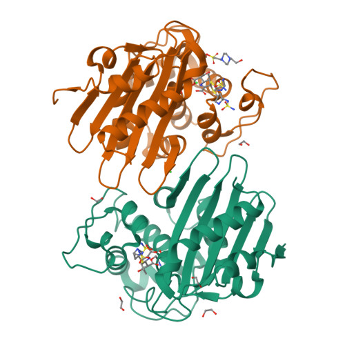
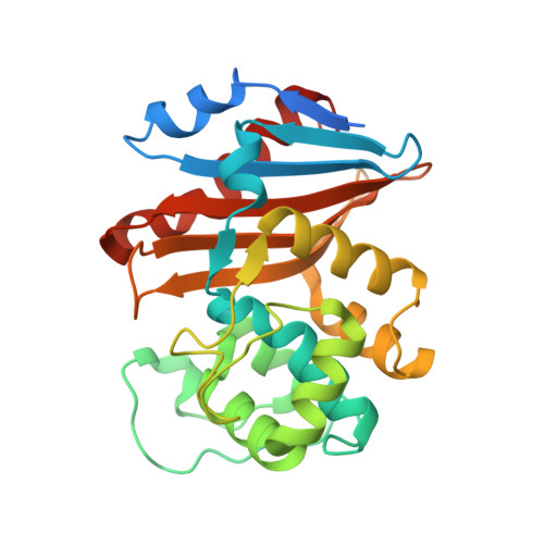
Structural Analysis of The OXA-48 Carbapenemase Bound to A "Poor" Carbapenem Substrate, Doripenem.

Papp-Wallace, K.M.Kumar, V.Zeiser, E.T.Becka, S.A.van den Akker, F.

(2019) Antibiotics (Basel) 8

  • DOI: https://doi.org/10.3390/antibiotics8030145
  • Primary Citation of Related Structures:  
    6PXX

  • PubMed Abstract: 

    Carbapenem-resistant Enterobacteriaceae are a significant threat to public health, and a major resistance determinant that promotes this phenotype is the production of the OXA-48 carbapenemase. The activity of OXA-48 towards carbapenems is a puzzling phenotype as its hydrolytic activity against doripenem is non-detectable. To probe the mechanistic basis for this observation, we determined the 1.5 Å resolution crystal structure of the deacylation deficient K73A variant of OXA-48 in complex with doripenem. Doripenem is observed in the Δ 1 R and Δ 1 S tautomeric states covalently attached to the catalytic S70 residue. Likely due to positioning of residue Y211, the carboxylate moiety of doripenem is making fewer hydrogen bonding/salt-bridge interactions with R250 compared to previously determined carbapenem OXA structures. Moreover, the hydroxyethyl side chain of doripenem is making van der Waals interactions with a key V120 residue, which likely affects the deacylation rate of doripenem. We hypothesize that positions V120 and Y211 play important roles in the carbapenemase profile of OXA-48. Herein, we provide insights for the further development of the carbapenem class of antibiotics that could render them less effective to hydrolysis by or even inhibit OXA carbapenemases.


  • Organizational Affiliation

    Department of Biochemistry, Case Western Reserve University, Cleveland, OH 44106, USA. krisztina.papp@va.gov.


Macromolecules
Find similar proteins by:  (by identity cutoff)  |  3D Structure
Entity ID: 1
MoleculeChains Sequence LengthOrganismDetailsImage
Beta-lactamase
A, B
265Klebsiella pneumoniaeMutation(s): 1 
Gene Names: bla OXA-48blaOXA-48KPE71T_00045
EC: 3.5.2.6
UniProt
Find proteins for Q6XEC0 (Klebsiella pneumoniae)
Explore Q6XEC0 
Go to UniProtKB:  Q6XEC0
Entity Groups  
Sequence Clusters30% Identity50% Identity70% Identity90% Identity95% Identity100% Identity
UniProt GroupQ6XEC0
Sequence Annotations
Expand
  • Reference Sequence
Small Molecules
Ligands 4 Unique
IDChains Name / Formula / InChI Key2D Diagram3D Interactions
5R7 (Subject of Investigation/LOI)
Query on 5R7

Download Ideal Coordinates CCD File 
C [auth A],
I [auth B]
(2~{S},3~{R})-3-methyl-2-[(2~{S},3~{R})-3-oxidanyl-1-oxidanylidene-butan-2-yl]-4-[(3~{S},5~{S})-5-[(sulfamoylamino)meth yl]pyrrolidin-3-yl]sulfanyl-3,4-dihydro-2~{H}-pyrrole-5-carboxylic acid
C15 H26 N4 O6 S2
RBKUPZPTQNSPAV-QZOCKBNKSA-N
EPE
Query on EPE

Download Ideal Coordinates CCD File 
J [auth B]4-(2-HYDROXYETHYL)-1-PIPERAZINE ETHANESULFONIC ACID
C8 H18 N2 O4 S
JKMHFZQWWAIEOD-UHFFFAOYSA-N
EDO
Query on EDO

Download Ideal Coordinates CCD File 
D [auth A],
E [auth A],
F [auth A],
G [auth A],
K [auth B]
1,2-ETHANEDIOL
C2 H6 O2
LYCAIKOWRPUZTN-UHFFFAOYSA-N
CL
Query on CL

Download Ideal Coordinates CCD File 
H [auth A],
L [auth B]
CHLORIDE ION
Cl
VEXZGXHMUGYJMC-UHFFFAOYSA-M
Experimental Data & Validation

Experimental Data

  • Method: X-RAY DIFFRACTION
  • Resolution: 1.50 Å
  • R-Value Free:  0.172 (Depositor), 0.171 (DCC) 
  • R-Value Work:  0.151 (Depositor), 0.151 (DCC) 
  • R-Value Observed: 0.152 (Depositor) 
Space Group: P 21 21 2
Unit Cell:
Length ( Å )Angle ( ˚ )
a = 105.77α = 90
b = 125.092β = 90
c = 45.12γ = 90
Software Package:
Software NamePurpose
REFMACrefinement
XDSdata reduction
Aimlessdata scaling
PDB_EXTRACTdata extraction
MOLREPphasing

Structure Validation

View Full Validation Report


Ligand Structure Quality Assessment 

Created with Raphaël 2.3.0Worse 01 BetterLigand structure goodness of fit to experimental dataBest fitted 5R7Click on this verticalbar to view details

Entry History 

Deposition Data

Revision History  (Full details and data files)

  • Version 1.0: 2019-10-09
    Type: Initial release
  • Version 1.1: 2024-11-20
    Changes: Data collection, Database references, Derived calculations, Structure summary