3HVJ | pdb_00003hvj

Rat catechol O-methyltransferase in complex with a catechol-type, N6-propyladenine-containing bisubstrate inhibitor


Experimental Data Snapshot

  • Method: X-RAY DIFFRACTION
  • Resolution: 1.79 Å
  • R-Value Free: 
    0.235 (Depositor), 0.240 (DCC) 
  • R-Value Work: 
    0.183 (Depositor), 0.190 (DCC) 
  • R-Value Observed: 
    0.186 (Depositor) 

wwPDB Validation   3D Report Full Report


Ligand Structure Quality Assessment 

Created with Raphaël 2.3.0Worse 01 BetterLigand structure goodness of fit to experimental dataBest fitted 705Click on this verticalbar to view details

This is version 1.4 of the entry. See complete history


Literature

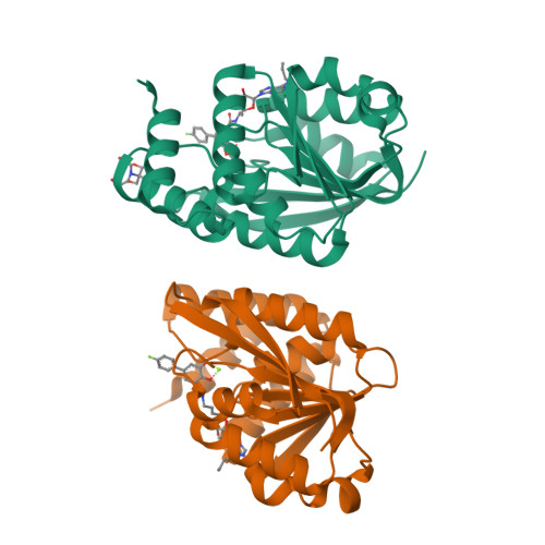
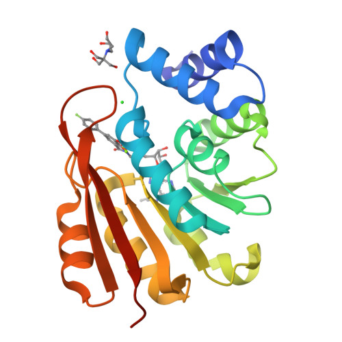
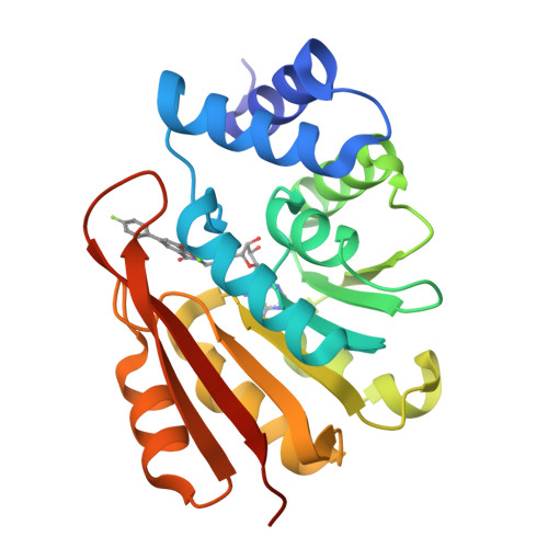
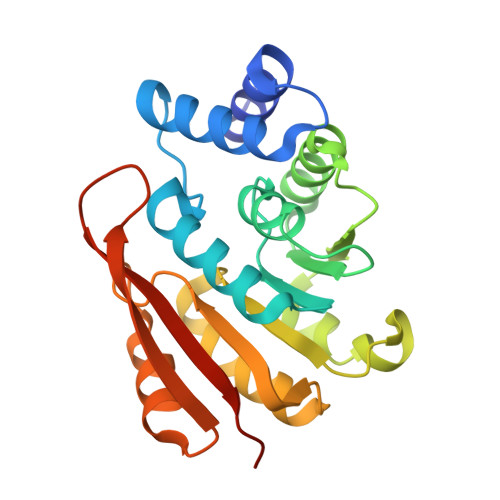
Molecular recognition at the active site of catechol-o-methyltransferase: energetically favorable replacement of a water molecule imported by a bisubstrate inhibitor.

Ellermann, M.Jakob-Roetne, R.Lerner, C.Borroni, E.Schlatter, D.Roth, D.Ehler, A.Rudolph, M.G.Diederich, F.

(2009) Angew Chem Int Ed Engl 48: 9092-9096


Macromolecules
Find similar proteins by:  (by identity cutoff)  |  3D Structure
Entity ID: 1
MoleculeChains Sequence LengthOrganismDetailsImage
Catechol O-methyltransferase
A, B
221Rattus norvegicusMutation(s): 0 
EC: 2.1.1.6
UniProt
Find proteins for P22734 (Rattus norvegicus)
Explore P22734 
Go to UniProtKB:  P22734
Entity Groups  
Sequence Clusters30% Identity50% Identity70% Identity90% Identity95% Identity100% Identity
UniProt GroupP22734
Sequence Annotations
Expand
  • Reference Sequence
Small Molecules
Ligands 4 Unique
IDChains Name / Formula / InChI Key2D Diagram3D Interactions
705
Query on 705

Download Ideal Coordinates CCD File 
C [auth A],
G [auth B]
N-[(E)-3-[(2R,3S,4R,5R)-3,4-dihydroxy-5-(6-propylaminopurin-9-yl)oxolan-2-yl]prop-2-enyl]-5-(4-fluorophenyl)-2,3-dihydroxy-benzamide
C28 H29 F N6 O6
ZTSZMLSDNBUFMA-WIRSHZJASA-N
BTB
Query on BTB

Download Ideal Coordinates CCD File 
F [auth A]2-[BIS-(2-HYDROXY-ETHYL)-AMINO]-2-HYDROXYMETHYL-PROPANE-1,3-DIOL
C8 H19 N O5
OWMVSZAMULFTJU-UHFFFAOYSA-N
CL
Query on CL

Download Ideal Coordinates CCD File 
E [auth A]CHLORIDE ION
Cl
VEXZGXHMUGYJMC-UHFFFAOYSA-M
MG
Query on MG

Download Ideal Coordinates CCD File 
D [auth A],
H [auth B]
MAGNESIUM ION
Mg
JLVVSXFLKOJNIY-UHFFFAOYSA-N
Experimental Data & Validation

Experimental Data

  • Method: X-RAY DIFFRACTION
  • Resolution: 1.79 Å
  • R-Value Free:  0.235 (Depositor), 0.240 (DCC) 
  • R-Value Work:  0.183 (Depositor), 0.190 (DCC) 
  • R-Value Observed: 0.186 (Depositor) 
Space Group: P 21 21 21
Unit Cell:
Length ( Å )Angle ( ˚ )
a = 55.346α = 90
b = 61.414β = 90
c = 132.5γ = 90
Software Package:
Software NamePurpose
COMOphasing
REFMACrefinement
XDSdata reduction
SADABSdata scaling

Structure Validation

View Full Validation Report


Ligand Structure Quality Assessment 

Created with Raphaël 2.3.0Worse 01 BetterLigand structure goodness of fit to experimental dataBest fitted 705Click on this verticalbar to view details

Entry History 

Deposition Data

Revision History  (Full details and data files)

  • Version 1.0: 2009-10-13
    Type: Initial release
  • Version 1.1: 2011-07-13
    Changes: Version format compliance
  • Version 1.2: 2012-08-08
    Changes: Database references, Structure summary
  • Version 1.3: 2017-11-01
    Changes: Refinement description
  • Version 1.4: 2024-03-20
    Changes: Data collection, Database references, Derived calculations