1TF2 | pdb_00001tf2

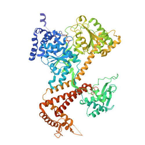
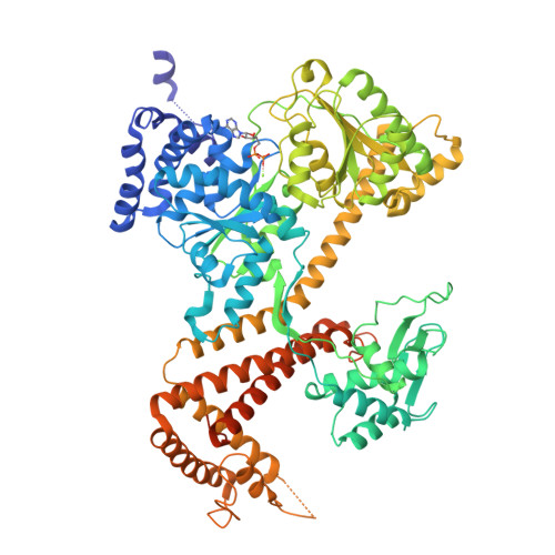
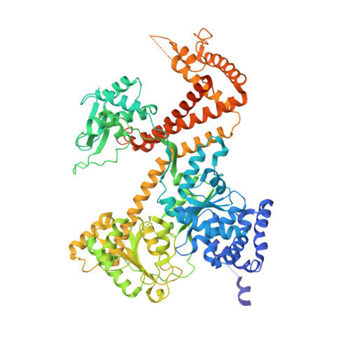
Crystal structure of SecA:ADP in an open conformation from Bacillus Subtilis


Experimental Data Snapshot

  • Method: X-RAY DIFFRACTION
  • Resolution: 2.90 Å
  • R-Value Free: 
    0.292 (Depositor), 0.290 (DCC) 
  • R-Value Work: 
    0.228 (Depositor), 0.230 (DCC) 
  • R-Value Observed: 
    0.228 (Depositor) 

Starting Model: experimental
View more details

wwPDB Validation   3D Report Full Report


Ligand Structure Quality Assessment 

Created with Raphaël 2.3.0Worse 01 BetterLigand structure goodness of fit to experimental dataBest fitted ADPClick on this verticalbar to view details

This is version 1.3 of the entry. See complete history


Literature

A large conformational change of the translocation ATPase SecA.

Osborne, A.R.Clemons Jr., W.M.Rapoport, T.A.

(2004) Proc Natl Acad Sci U S A 101: 10937-10942

  • DOI: https://doi.org/10.1073/pnas.0401742101
  • Primary Citation of Related Structures:  
    1TF2, 1TF5

  • PubMed Abstract: 

    The ATPase SecA mediates the posttranslational translocation of a wide range of polypeptide substrates through the SecY channel in the cytoplasmic membrane of bacteria. We have determined the crystal structure of a monomeric form of Bacillus subtilis SecA at a 2.2-A resolution. A comparison with the previously determined structures of SecA reveals a nucleotide-independent, large conformational change that opens a deep groove similar to that in other proteins that interact with diverse polypeptides. We propose that the open form of SecA represents an activated state.


  • Organizational Affiliation

    Department of Cell Biology, Harvard Medical School, Boston, MA 02115, USA.


Macromolecules
Find similar proteins by:  (by identity cutoff)  |  3D Structure
Entity ID: 1
MoleculeChains Sequence LengthOrganismDetailsImage
Preprotein translocase secA subunit844Bacillus subtilisMutation(s): 0 
Gene Names: SECADIV+BSU35300
EC: 7.4.2.8
UniProt
Find proteins for P28366 (Bacillus subtilis (strain 168))
Explore P28366 
Go to UniProtKB:  P28366
Entity Groups  
Sequence Clusters30% Identity50% Identity70% Identity90% Identity95% Identity100% Identity
UniProt GroupP28366
Sequence Annotations
Expand
  • Reference Sequence
Experimental Data & Validation

Experimental Data

  • Method: X-RAY DIFFRACTION
  • Resolution: 2.90 Å
  • R-Value Free:  0.292 (Depositor), 0.290 (DCC) 
  • R-Value Work:  0.228 (Depositor), 0.230 (DCC) 
  • R-Value Observed: 0.228 (Depositor) 
Space Group: C 1 2 1
Unit Cell:
Length ( Å )Angle ( ˚ )
a = 149.232α = 90
b = 107.189β = 94.96
c = 72.054γ = 90
Software Package:
Software NamePurpose
CNSrefinement
HKL-2000data reduction
SCALEPACKdata scaling
AMoREphasing

Structure Validation

View Full Validation Report


Ligand Structure Quality Assessment 

Created with Raphaël 2.3.0Worse 01 BetterLigand structure goodness of fit to experimental dataBest fitted ADPClick on this verticalbar to view details

Entry History 

Deposition Data

Revision History  (Full details and data files)

  • Version 1.0: 2004-08-03
    Type: Initial release
  • Version 1.1: 2008-04-30
    Changes: Version format compliance
  • Version 1.2: 2011-07-13
    Changes: Version format compliance
  • Version 1.3: 2023-08-23
    Changes: Data collection, Database references, Derived calculations, Refinement description