1LK2 | pdb_00001lk2

1.35A crystal structure of H-2Kb complexed with the GNYSFYAL peptide


Experimental Data Snapshot

  • Method: X-RAY DIFFRACTION
  • Resolution: 1.35 Å
  • R-Value Free: 
    0.164 (Depositor), 0.243 (DCC) 
  • R-Value Work: 
    0.149 (Depositor), 0.234 (DCC) 
  • R-Value Observed: 
    0.150 (Depositor) 

wwPDB Validation   3D Report Full Report


Ligand Structure Quality Assessment 

Created with Raphaël 2.3.0Worse 01 BetterLigand structure goodness of fit to experimental dataBest fitted NAGClick on this verticalbar to view details

This is version 2.1 of the entry. See complete history


Literature

A peptide that antagonizes TCR-mediated reactions with both syngeneic and allogeneic agonists: functional and structural aspects.

Rudolph, M.G.Shen, L.Q.Lamontagne, S.A.Luz, J.G.Delaney, J.R.Ge, Q.Cho, B.K.Palliser, D.McKinley, C.A.Chen, J.Wilson, I.A.Eisen, H.N.

(2004) J Immunol 172: 2994-3002

  • DOI: https://doi.org/10.4049/jimmunol.172.5.2994
  • Primary Citation of Related Structures:  
    1LK2

  • PubMed Abstract: 

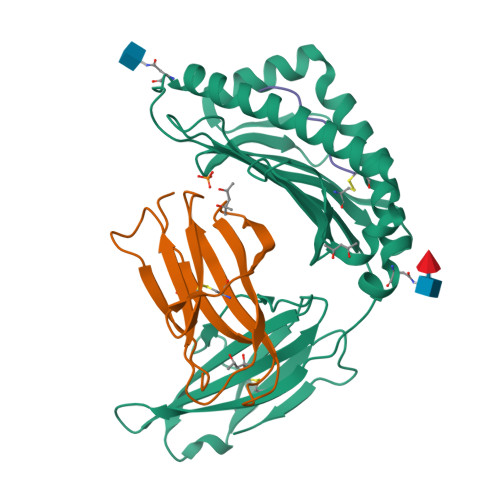
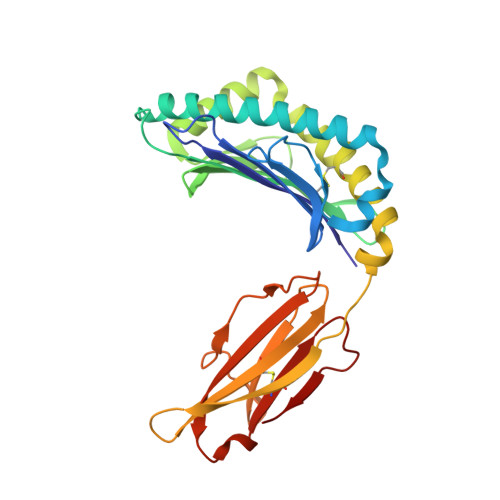
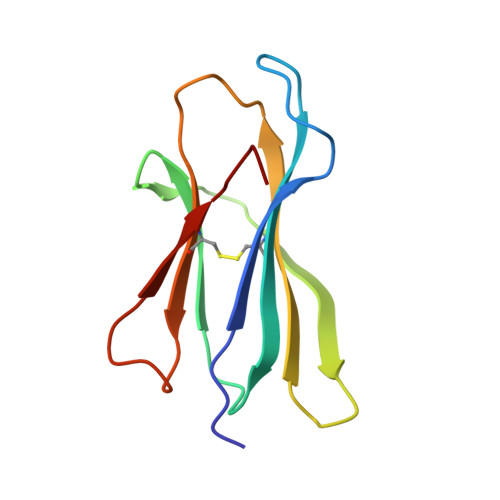
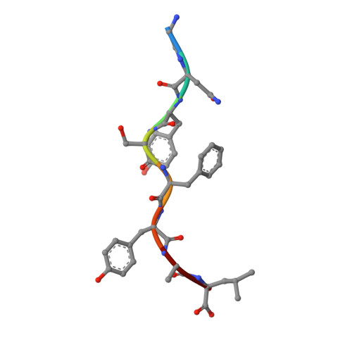
    We identify and consider some characteristics of a peptide antagonist for the Ag-specific receptor on 2C cells (the 2C TCR). The peptide, GNYSFYAL (called GNY), binds to H-2K(b), and a very high-resolution crystal structure of the GNY-K(b) complex at 1.35 A is described. Although the GNY peptide does not bind to L(d), the potency of GNY-K(b) as an antagonist is evident from its ability to specifically inhibit 2C TCR-mediated reactions to an allogenic agonist complex (QLSPFPFDL-L(d)), as well as to a syngeneic agonist complex (SIYRYYGL-K(b)). The crystal structure and the activities of alanine-substituted peptide variants point to the properties of the peptide P4 side chain and the conformation of the Tyr-P6 side chain as the structural determinants of GNYSFYAL antagonist activity.


  • Organizational Affiliation

    Center for Cancer Research and Department of Biology, Massachusetts Institute of Technology, Cambridge, MA 02139, USA. Markus.Rudolph@bio.uni-goettingen.de


Macromolecules
Find similar proteins by:  (by identity cutoff)  |  3D Structure
Entity ID: 1
MoleculeChains Sequence LengthOrganismDetailsImage
H-2 CLASS I HISTOCOMPATIBILITY ANTIGEN, K-B ALPHA CHAIN274Mus musculusMutation(s): 0 
UniProt
Find proteins for P01901 (Mus musculus)
Explore P01901 
Go to UniProtKB:  P01901
Entity Groups  
Sequence Clusters30% Identity50% Identity70% Identity90% Identity95% Identity100% Identity
UniProt GroupP01901
Glycosylation
Glycosylation Sites: 2Go to GlyGen: P01901-1
Sequence Annotations
Expand
  • Reference Sequence
Find similar proteins by:  (by identity cutoff)  |  3D Structure
Entity ID: 2
MoleculeChains Sequence LengthOrganismDetailsImage
Beta-2-microglobulin99Mus musculusMutation(s): 0 
UniProt & NIH Common Fund Data Resources
Find proteins for P01887 (Mus musculus)
Explore P01887 
Go to UniProtKB:  P01887
IMPC:  MGI:88127
Entity Groups  
Sequence Clusters30% Identity50% Identity70% Identity90% Identity95% Identity100% Identity
UniProt GroupP01887
Sequence Annotations
Expand
  • Reference Sequence

Find similar proteins by:  Sequence   |   3D Structure  

Entity ID: 3
MoleculeChains Sequence LengthOrganismDetailsImage
INSULIN RECEPTOR, BETA-SUBUNITC [auth P]8N/AMutation(s): 0 
EC: 2.7.10.1
UniProt
Find proteins for P15208 (Mus musculus)
Explore P15208 
Go to UniProtKB:  P15208
Entity Groups  
UniProt GroupP15208
Sequence Annotations
Expand
  • Reference Sequence
Oligosaccharides

Help

Entity ID: 4
MoleculeChains Length2D Diagram Glycosylation3D Interactions
alpha-L-fucopyranose-(1-6)-2-acetamido-2-deoxy-beta-D-glucopyranoseD [auth C]2N-Glycosylation
Glycosylation Resources
GlyTouCan:  G86851RC
GlyCosmos:  G86851RC
GlyGen:  G86851RC
Small Molecules
Ligands 4 Unique
IDChains Name / Formula / InChI Key2D Diagram3D Interactions
NAG
Query on NAG

Download Ideal Coordinates CCD File 
E [auth A]2-acetamido-2-deoxy-beta-D-glucopyranose
C8 H15 N O6
OVRNDRQMDRJTHS-FMDGEEDCSA-N
MRD
Query on MRD

Download Ideal Coordinates CCD File 
H [auth B](4R)-2-METHYLPENTANE-2,4-DIOL
C6 H14 O2
SVTBMSDMJJWYQN-RXMQYKEDSA-N
MPD
Query on MPD

Download Ideal Coordinates CCD File 
G [auth A],
I [auth B]
(4S)-2-METHYL-2,4-PENTANEDIOL
C6 H14 O2
SVTBMSDMJJWYQN-YFKPBYRVSA-N
PO4
Query on PO4

Download Ideal Coordinates CCD File 
F [auth A]PHOSPHATE ION
O4 P
NBIIXXVUZAFLBC-UHFFFAOYSA-K
Experimental Data & Validation

Experimental Data

  • Method: X-RAY DIFFRACTION
  • Resolution: 1.35 Å
  • R-Value Free:  0.164 (Depositor), 0.243 (DCC) 
  • R-Value Work:  0.149 (Depositor), 0.234 (DCC) 
  • R-Value Observed: 0.150 (Depositor) 
Space Group: P 21 21 2
Unit Cell:
Length ( Å )Angle ( ˚ )
a = 135.549α = 90
b = 87.65β = 90
c = 45.118γ = 90
Software Package:
Software NamePurpose
REFMACrefinement
SCALEPACKdata scaling
CNSrefinement
DENZOdata reduction
CNSphasing

Structure Validation

View Full Validation Report


Ligand Structure Quality Assessment 

Created with Raphaël 2.3.0Worse 01 BetterLigand structure goodness of fit to experimental dataBest fitted NAGClick on this verticalbar to view details

Entry History 

Deposition Data

Revision History  (Full details and data files)

  • Version 1.0: 2003-11-11
    Type: Initial release
  • Version 1.1: 2008-04-28
    Changes: Version format compliance
  • Version 1.2: 2011-07-13
    Changes: Non-polymer description, Version format compliance
  • Version 1.3: 2019-07-24
    Changes: Data collection, Derived calculations, Refinement description
  • Version 2.0: 2020-07-29
    Type: Remediation
    Reason: Carbohydrate remediation
    Changes: Advisory, Atomic model, Data collection, Derived calculations, Structure summary
  • Version 2.1: 2024-11-13
    Changes: Data collection, Database references, Derived calculations, Structure summary