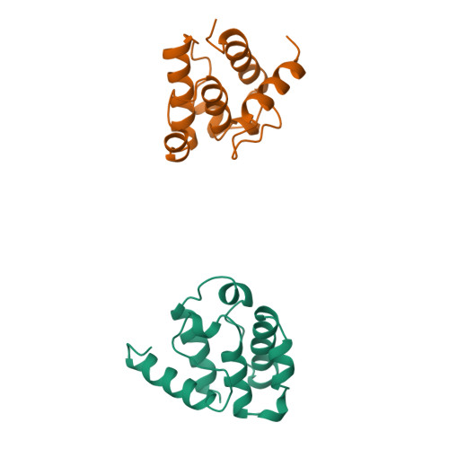
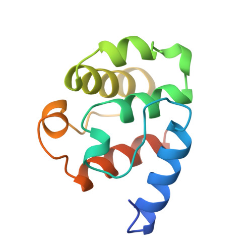
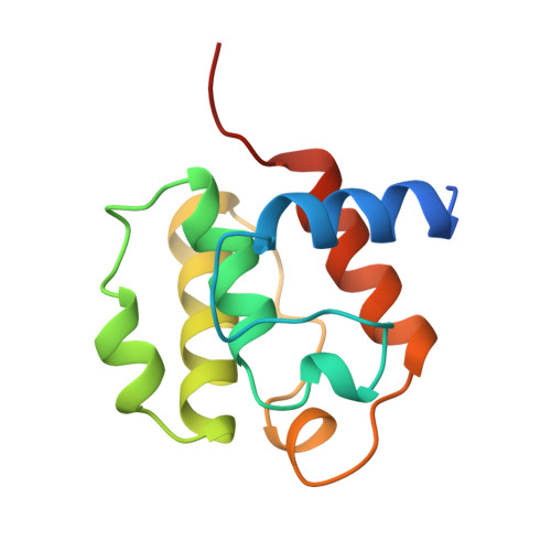
The 2.0 A structure of the second calponin homology domain from the actin-binding region of the dystrophin homologue utrophin.
Keep, N.H., Norwood, F.L., Moores, C.A., Winder, S.J., Kendrick-Jones, J.(1999) J Mol Biology 285: 1257-1264
- PubMed: 9887274
- DOI: https://doi.org/10.1006/jmbi.1998.2406
- Primary Citation of Related Structures:
1BHD - PubMed Abstract:
Utrophin is a close homologue of dystrophin, the protein defective in Duchenne muscular dystrophy. Like dystrophin, it is composed of three regions: an N-terminal region that binds actin filaments, a large central region with triple coiled-coil repeats, and a C-terminal region that interacts with components in the dystroglycan protein complex at the plasma membrane. The N-terminal actin-binding region consists of two calponin homology domains and is related to the actin-binding domains of a superfamily of proteins including alpha-actinin, spectrin and fimbrin. Here, we present the 2.0 A structure of the second calponin homology domain of utrophin solved by X-ray crystallography, and compare it to the other calponin homology domains previously determined from spectrin and fimbrin.
Organizational Affiliation:
MRC Laboratory of Molecular Biology, Hills Road, Cambridge, CB2 2QH, UK. n.keep@mail.cryst.bbk.ac.uk


