1B23 | pdb_00001b23

E. coli cysteinyl-tRNA and T. aquaticus elongation factor EF-TU:GTP ternary complex


Experimental Data Snapshot

  • Method: X-RAY DIFFRACTION
  • Resolution: 2.60 Å
  • R-Value Free: 
    0.263 (Depositor), 0.260 (DCC) 
  • R-Value Work: 
    0.206 (Depositor), 0.210 (DCC) 
  • R-Value Observed: 
    0.206 (Depositor) 

Starting Model: experimental
View more details

wwPDB Validation   3D Report Full Report


Ligand Structure Quality Assessment 

Created with Raphaël 2.3.0Worse 01 BetterLigand structure goodness of fit to experimental dataBest fitted GNPClick on this verticalbar to view details

This is version 2.1 of the entry. See complete history


Literature

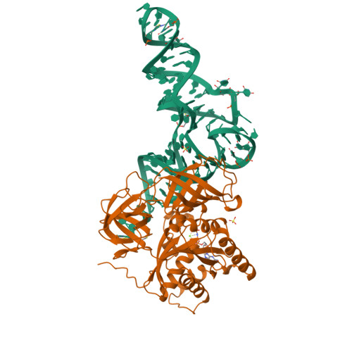
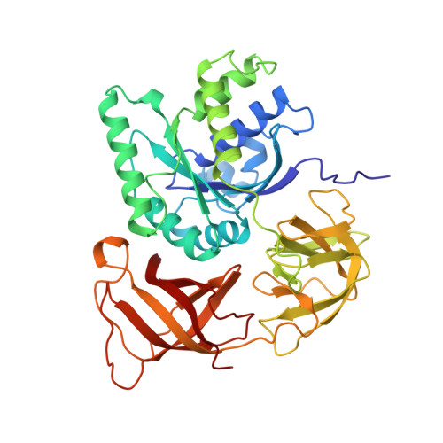
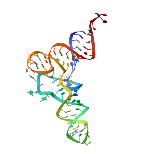
The crystal structure of Cys-tRNACys-EF-Tu-GDPNP reveals general and specific features in the ternary complex and in tRNA.

Nissen, P.Thirup, S.Kjeldgaard, M.Nyborg, J.

(1999) Structure 7: 143-156

  • DOI: https://doi.org/10.1016/s0969-2126(99)80021-5
  • Primary Citation of Related Structures:  
    1B23

  • PubMed Abstract: 

    . The translation elongation factor EF-Tu in its GTP-bound state forms a ternary complex with any aminoacylated tRNA (aa-tRNA), except initiator tRNA and selenocysteinyl-tRNA. This complex delivers aa-tRNA to the ribosomal A site during the elongation cycle of translation. The crystal structure of the yeast Phe-tRNAPhe ternary complex with Thermus aquaticus EF-Tu-GDPNP (Phe-TC) has previously been determined as one representative of this general yet highly discriminating complex formation. The ternary complex of Escherichia coli Cys-tRNACys and T. aquaticus EF-Tu-GDPNP (Cys-TC) has been solved and refined at 2.6 degrees resolution. Conserved and variable features of the aa-tRNA recognition and binding by EF-Tu-GTP have been revealed by comparison with the Phe-TC structure. New tertiary interactions are observed in the tRNACys structure. A 'kissing complex' is observed in the very close crystal packing arrangement. The recognition of Cys-tRNACys by EF-Tu-GDPNP is restricted to the aa-tRNA motif previously identified in Phe-TC and consists of the aminoacylated 3' end, the phosphorylated 5' end and one side of the acceptor stem and T stem. The aminoacyl bond is recognized somewhat differently, yet by the same primary motif in EF-Tu, which suggests that EF-Tu adapts to subtle variations in this moiety among all aa-tRNAs. New tertiary interactions revealed by the Cys-tRNACys structure, such as a protonated C16:C59 pyrimidine pair, a G15:G48 'Levitt pair' and an s4U8:A14:A46 base triple add to the generic understanding of tRNA structure from sequence. The structure of the 'kissing complex' shows a quasicontinuous helix with a distinct shape determined by the number of base pairs.


  • Organizational Affiliation

    Institute of Molecular and Structural Biology, Aarhus University, Gustav Wieds Vej 10, C DK 8000 Aarhus C, Denmark.


Macromolecules

Find similar proteins by:  (by identity cutoff)  |  3D Structure
Entity ID: 2
MoleculeChains Sequence LengthOrganismDetailsImage
ELONGATION FACTOR TUB [auth P]405Thermus aquaticusMutation(s): 0 
Gene Names: TUFA
UniProt
Find proteins for Q01698 (Thermus aquaticus)
Explore Q01698 
Go to UniProtKB:  Q01698
Entity Groups  
Sequence Clusters30% Identity50% Identity70% Identity90% Identity95% Identity100% Identity
UniProt GroupQ01698
Sequence Annotations
Expand
  • Reference Sequence
Small Molecules
Ligands 4 Unique
IDChains Name / Formula / InChI Key2D Diagram3D Interactions
GNP
Query on GNP

Download Ideal Coordinates CCD File 
I [auth P]PHOSPHOAMINOPHOSPHONIC ACID-GUANYLATE ESTER
C10 H17 N6 O13 P3
UQABYHGXWYXDTK-UUOKFMHZSA-N
CYS
Query on CYS

Download Ideal Coordinates CCD File 
E [auth R]CYSTEINE
C3 H7 N O2 S
XUJNEKJLAYXESH-REOHCLBHSA-N
SO4
Query on SO4

Download Ideal Coordinates CCD File 
G [auth P],
H [auth P]
SULFATE ION
O4 S
QAOWNCQODCNURD-UHFFFAOYSA-L
MG
Query on MG

Download Ideal Coordinates CCD File 
C [auth R],
D [auth R],
F [auth P]
MAGNESIUM ION
Mg
JLVVSXFLKOJNIY-UHFFFAOYSA-N
Experimental Data & Validation

Experimental Data

  • Method: X-RAY DIFFRACTION
  • Resolution: 2.60 Å
  • R-Value Free:  0.263 (Depositor), 0.260 (DCC) 
  • R-Value Work:  0.206 (Depositor), 0.210 (DCC) 
  • R-Value Observed: 0.206 (Depositor) 
Space Group: F 2 2 2
Unit Cell:
Length ( Å )Angle ( ˚ )
a = 126.75α = 90
b = 132.98β = 90
c = 154.88γ = 90
Software Package:
Software NamePurpose
AMoREphasing
X-PLORrefinement
DENZOdata reduction
SCALEPACKdata scaling

Structure Validation

View Full Validation Report


Ligand Structure Quality Assessment 

Created with Raphaël 2.3.0Worse 01 BetterLigand structure goodness of fit to experimental dataBest fitted GNPClick on this verticalbar to view details

Entry History 

Deposition Data

Revision History  (Full details and data files)

  • Version 1.0: 1998-12-07
    Type: Initial release
  • Version 1.1: 2008-04-26
    Changes: Version format compliance
  • Version 1.2: 2011-07-13
    Changes: Version format compliance
  • Version 1.3: 2011-11-16
    Changes: Atomic model
  • Version 2.0: 2019-07-03
    Changes: Advisory, Data collection, Derived calculations, Non-polymer description, Structure summary
  • Version 2.1: 2023-08-09
    Changes: Advisory, Data collection, Database references, Derived calculations, Refinement description