9FMM | pdb_00009fmm

Structure of human ACE2 in complex with a fluorinated small molecule inhibitor


Experimental Data Snapshot

  • Method: X-RAY DIFFRACTION
  • Resolution: 2.50 Å
  • R-Value Free: 
    0.265 (Depositor), 0.270 (DCC) 
  • R-Value Work: 
    0.213 (Depositor), 0.210 (DCC) 
  • R-Value Observed: 
    0.215 (Depositor) 

Starting Model: in silico
View more details

wwPDB Validation   3D Report Full Report


Ligand Structure Quality Assessment 

Created with Raphaël 2.3.0Worse 01 BetterLigand structure goodness of fit to experimental dataBest fitted A1IDXClick on this verticalbar to view details

This is version 1.3 of the entry. See complete history


Literature

Development of radiofluorinated MLN-4760 derivatives for PET imaging of the SARS-CoV-2 entry receptor ACE2.

Wang, J.Beyer, D.Vaccarin, C.He, Y.Tanriver, M.Benoit, R.Deupi, X.Mu, L.Bode, J.W.Schibli, R.Muller, C.

(2024) Eur J Nucl Med Mol Imaging 52: 9-21

  • DOI: https://doi.org/10.1007/s00259-024-06831-6
  • Primary Citation of Related Structures:  
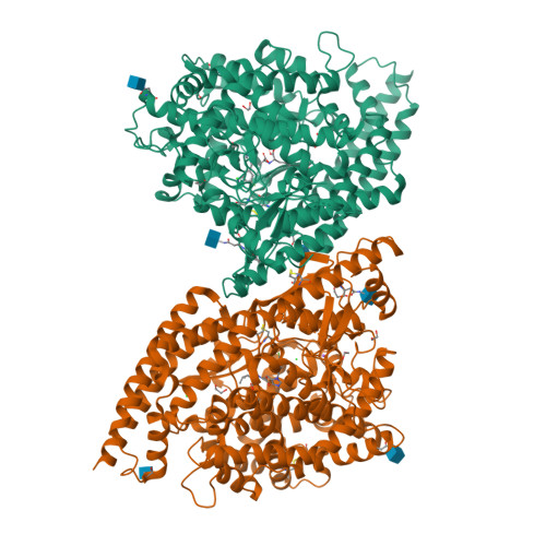
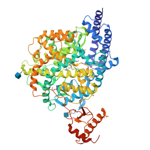
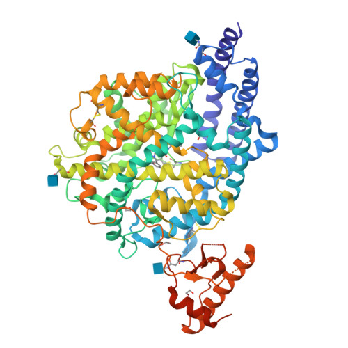
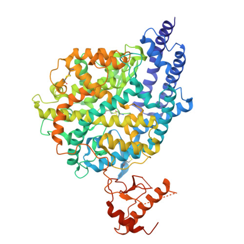
    9FMM

  • PubMed Abstract: 

    The angiotensin converting enzyme 2 (ACE2) plays a regulatory role in the cardiovascular system and serves SARS-CoV-2 as an entry receptor. The aim of this study was to synthesize and evaluate radiofluorinated derivatives of the ACE2 inhibitor MLN-4760. [ 18 F]F-MLN-4760 and [ 18 F]F-Aza-MLN-4760 were demonstrated to be suitable for non-invasive imaging of ACE2, potentially enabling a better understanding of its expression dynamics. Computational molecular modeling, based on the structures of human ACE2 (hACE2) and mouse ACE2 (mACE2), revealed that the ACE2-binding modes of F-MLN-4760 and F-Aza-MLN-4760 were similar to that of MLN-4760. Co-crystallization of the hACE2/F-MLN-4760 protein complex was performed for confirmation. Displacement experiments using [ 3 H]MLN-4760 enabled the determination of the binding affinities of the synthesized F-MLN-4760 and F-Aza-MLN-4760 to hACE2 expressed in HEK-ACE2 cells. Aryl trimethylstannane-based and pyridine-based radiofluorination precursors were synthesized and used for the preparation of the respective radiotracers. [ 18 F]F-MLN-4760 and [ 18 F]F-Aza-MLN-4760 were evaluated with regard to the uptake in HEK-ACE2 and HEK-ACE cells and in vitro binding to tissue sections of HEK-ACE2 xenografts and normal organs of mice. Biodistribution and PET/CT imaging studies of [ 18 F]F-MLN-4760 and [ 18 F]F-Aza-MLN-4760 were performed using HEK-ACE2 and HEK-ACE xenografted nude mice. Crystallography data revealed an equal hACE2-binding mode for F-MLN-4760 as previously found for MLN-4760. Moreover, computer-based modeling indicated that similar binding to hACE2 and mACE2 holds true for both, F-MLN-4760 and F-Aza-MLN-4760, as is the case for MLN-4760. The IC 50 values were three-fold and seven-fold higher for F-MLN-4760 and F-Aza-MLN-4760, respectively, than for MLN-4760. [ 18 F]F-MLN-4760 and [ 18 F]F-Aza-MLN-4760 were obtained in 1.4 ± 0.3 GBq and 0.5 ± 0.1 GBq activity with > 99% radiochemical purity in a 5.3% and 1.2% radiochemical yield, respectively. Uptake in HEK-ACE2 cells was higher for [ 18 F]F-MLN-4760 (67 ± 9%) than for [ 18 F]F-Aza-MLN-4760 (37 ± 8%) after 3-h incubation while negligible uptake was seen in HEK-ACE cells (< 0.3%). [ 18 F]F-MLN-4760 and [ 18 F]F-Aza-MLN-4760 accumulated specifically in HEK-ACE2 xenografts of mice (13 ± 2% IA/g and 15 ± 2% IA/g at 1 h p.i.) with almost no uptake observed in HEK-ACE xenografts (< 0.3% IA/g). This was confirmed by PET/CT imaging, which also visualized unspecific accumulation in the gall bladder and intestinal tract. Both radiotracers showed specific and selective binding to ACE2 in vitro and in vivo. [ 18 F]F-MLN-4760 was, however, obtained in higher yields and the ACE2-binding affinity was superior over that of [ 18 F]F-Aza-MLN-4760. [ 18 F]F-MLN-4760 would, thus, be the candidate of choice for further development in view of its use for PET imaging of ACE2.


  • Organizational Affiliation

    Laboratory of Organic Chemistry, Department of Chemistry and Applied Biosciences, ETH Zurich, Zurich, 8093, Switzerland.


Macromolecules
Find similar proteins by:  (by identity cutoff)  |  3D Structure
Entity ID: 1
MoleculeChains Sequence LengthOrganismDetailsImage
Angiotensin-converting enzyme 2
A, B
730Homo sapiensMutation(s): 0 
Gene Names: ACE2UNQ868/PRO1885
EC: 3.4.17.23 (PDB Primary Data), 3.4.17 (PDB Primary Data)
UniProt & NIH Common Fund Data Resources
Find proteins for Q9BYF1 (Homo sapiens)
Explore Q9BYF1 
Go to UniProtKB:  Q9BYF1
PHAROS:  Q9BYF1
GTEx:  ENSG00000130234 
Entity Groups  
Sequence Clusters30% Identity50% Identity70% Identity90% Identity95% Identity100% Identity
UniProt GroupQ9BYF1
Glycosylation
Glycosylation Sites: 3Go to GlyGen: Q9BYF1-1
Sequence Annotations
Expand
  • Reference Sequence
Small Molecules
Ligands 6 Unique
IDChains Name / Formula / InChI Key2D Diagram3D Interactions
A1IDX (Subject of Investigation/LOI)
Query on A1IDX

Download Ideal Coordinates CCD File 
C [auth A],
P [auth B]
(2~{S})-2-[[(2~{S})-3-[3-[(3-chloranyl-5-fluoranyl-phenyl)methyl]imidazol-4-yl]-1-oxidanyl-1-oxidanylidene-propan-2-yl]amino]-4-methyl-pentanoic acid
C19 H23 Cl F N3 O4
OOQXYGMWUYCKSJ-IRXDYDNUSA-N
NAG
Query on NAG

Download Ideal Coordinates CCD File 
D [auth A]
E [auth A]
G [auth A]
O [auth B]
Q [auth B]
2-acetamido-2-deoxy-beta-D-glucopyranose
C8 H15 N O6
OVRNDRQMDRJTHS-FMDGEEDCSA-N
ZN
Query on ZN

Download Ideal Coordinates CCD File 
N [auth A],
Z [auth B]
ZINC ION
Zn
PTFCDOFLOPIGGS-UHFFFAOYSA-N
EDO
Query on EDO

Download Ideal Coordinates CCD File 
F [auth A]
H [auth A]
I [auth A]
J [auth A]
K [auth A]
1,2-ETHANEDIOL
C2 H6 O2
LYCAIKOWRPUZTN-UHFFFAOYSA-N
CL
Query on CL

Download Ideal Coordinates CCD File 
L [auth A],
W [auth B],
X [auth B]
CHLORIDE ION
Cl
VEXZGXHMUGYJMC-UHFFFAOYSA-M
NA
Query on NA

Download Ideal Coordinates CCD File 
M [auth A],
Y [auth B]
SODIUM ION
Na
FKNQFGJONOIPTF-UHFFFAOYSA-N
Experimental Data & Validation

Experimental Data

  • Method: X-RAY DIFFRACTION
  • Resolution: 2.50 Å
  • R-Value Free:  0.265 (Depositor), 0.270 (DCC) 
  • R-Value Work:  0.213 (Depositor), 0.210 (DCC) 
  • R-Value Observed: 0.215 (Depositor) 
Space Group: P 1 21 1
Unit Cell:
Length ( Å )Angle ( ˚ )
a = 99.3α = 90
b = 82.9β = 104.2
c = 105.5γ = 90
Software Package:
Software NamePurpose
PHENIXrefinement
PHENIXrefinement
XDSdata reduction
XSCALEdata scaling
PHASERphasing

Structure Validation

View Full Validation Report


Ligand Structure Quality Assessment 

Created with Raphaël 2.3.0Worse 01 BetterLigand structure goodness of fit to experimental dataBest fitted A1IDXClick on this verticalbar to view details

Entry History & Funding Information

Deposition Data


Funding OrganizationLocationGrant Number
Other privatePromedica 1401/M
Swiss National Science FoundationSwitzerlandCRSK-3_190414
Swiss National Science FoundationSwitzerland198274

Revision History  (Full details and data files)

  • Version 1.0: 2024-07-17
    Type: Initial release
  • Version 1.1: 2024-08-14
    Changes: Database references
  • Version 1.2: 2024-10-16
    Changes: Structure summary
  • Version 1.3: 2024-12-04
    Changes: Database references