7ZQZ

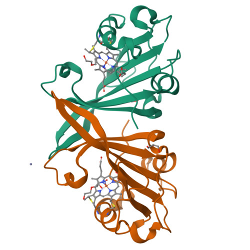
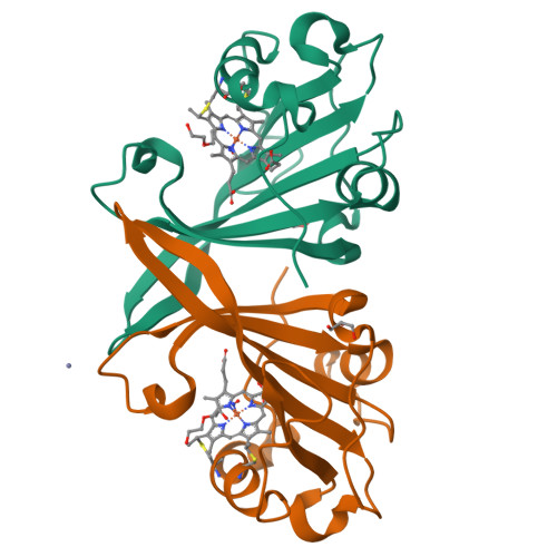
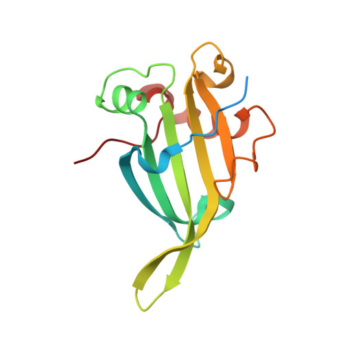
F61V Cytochrome c prime beta from Methylococcus capsulatus (Bath): NO complex


Experimental Data Snapshot

  • Method: X-RAY DIFFRACTION
  • Resolution: 1.68 Å
  • R-Value Free: 0.202 
  • R-Value Work: 0.178 
  • R-Value Observed: 0.179 

Starting Model: experimental
View more details

wwPDB Validation   3D Report Full Report


This is version 1.3 of the entry. See complete history


Literature

A heme pocket aromatic quadrupole modulates gas binding to cytochrome c'-beta : Implications for NO sensors.

Adams, H.R.Svistunenko, D.A.Wilson, M.T.Fujii, S.Strange, R.W.Hardy, Z.A.Vazquez, P.A.Dabritz, T.Streblow, G.J.Andrew, C.R.Hough, M.A.

(2023) J Biol Chem 299: 104742-104742

  • DOI: https://doi.org/10.1016/j.jbc.2023.104742
  • Primary Citation of Related Structures:  
    7ZPS, 7ZQZ, 7ZRW, 7ZRX, 7ZS4, 7ZSV, 7ZSW, 7ZSX, 7ZTI, 7ZVZ

  • PubMed Abstract: 

    The structural basis by which gas-binding heme proteins control their interactions with NO, CO, and O 2 is fundamental to enzymology, biotechnology, and human health. Cytochromes c' (cyts c') are a group of putative NO-binding heme proteins that fall into two families: the well-characterized four alpha helix bundle fold (cyts c'-α) and an unrelated family with a large beta-sheet fold (cyts c'-β) resembling that of cytochromes P460. A recent structure of cyt c'-β from Methylococcus capsulatus Bath revealed two heme pocket phenylalanine residues (Phe 32 and Phe 61) positioned near the distal gas-binding site. This feature, dubbed the "Phe cap," is highly conserved within the sequences of other cyts c'-β but is absent in their close homologs, the hydroxylamine-oxidizing cytochromes P460, although some do contain a single Phe residue. Here, we report an integrated structural, spectroscopic, and kinetic characterization of cyt c'-β from Methylococcus capsulatus Bath complexes with diatomic gases, focusing on the interaction of the Phe cap with NO and CO. Significantly, crystallographic and resonance Raman data show that orientation of the electron-rich aromatic ring face of Phe 32 toward distally bound NO or CO is associated with weakened backbonding and higher off rates. Moreover, we propose that an aromatic quadrupole also contributes to the unusually weak backbonding reported for some heme-based gas sensors, including the mammalian NO sensor, soluble guanylate cyclase. Collectively, this study sheds light on the influence of highly conserved distal Phe residues on heme-gas complexes of cytochrome c'-β, including the potential for aromatic quadrupoles to modulate NO and CO binding in other heme proteins.


  • Organizational Affiliation

    School of Life Sciences, University of Essex, Colchester, Essex, United Kingdom.


Macromolecules
Find similar proteins by:  (by identity cutoff)  |  3D Structure
Entity ID: 1
MoleculeChains Sequence LengthOrganismDetailsImage
Cytochrome c
A, B
158Methylococcus capsulatus str. BathMutation(s): 1 
Gene Names: ccpMCA2394
UniProt
Find proteins for G1UBD5 (Methylococcus capsulatus (strain ATCC 33009 / NCIMB 11132 / Bath))
Explore G1UBD5 
Go to UniProtKB:  G1UBD5
Entity Groups  
Sequence Clusters30% Identity50% Identity70% Identity90% Identity95% Identity100% Identity
UniProt GroupG1UBD5
Sequence Annotations
Expand
  • Reference Sequence
Small Molecules
Ligands 5 Unique
IDChains Name / Formula / InChI Key2D Diagram3D Interactions
HEC
Query on HEC

Download Ideal Coordinates CCD File 
D [auth A],
G [auth B]
HEME C
C34 H34 Fe N4 O4
HXQIYSLZKNYNMH-LJNAALQVSA-N
PEG
Query on PEG

Download Ideal Coordinates CCD File 
F [auth A],
K [auth B]
DI(HYDROXYETHYL)ETHER
C4 H10 O3
MTHSVFCYNBDYFN-UHFFFAOYSA-N
GOL
Query on GOL

Download Ideal Coordinates CCD File 
H [auth B],
I [auth B]
GLYCEROL
C3 H8 O3
PEDCQBHIVMGVHV-UHFFFAOYSA-N
ZN
Query on ZN

Download Ideal Coordinates CCD File 
C [auth A]ZINC ION
Zn
PTFCDOFLOPIGGS-UHFFFAOYSA-N
NO (Subject of Investigation/LOI)
Query on NO

Download Ideal Coordinates CCD File 
E [auth A],
J [auth B]
NITRIC OXIDE
N O
ODUCDPQEXGNKDN-UHFFFAOYSA-N
Experimental Data & Validation

Experimental Data

  • Method: X-RAY DIFFRACTION
  • Resolution: 1.68 Å
  • R-Value Free: 0.202 
  • R-Value Work: 0.178 
  • R-Value Observed: 0.179 
  • Space Group: P 21 3
Unit Cell:
Length ( Å )Angle ( ˚ )
a = 106.236α = 90
b = 106.236β = 90
c = 106.236γ = 90
Software Package:
Software NamePurpose
Aimlessdata scaling
REFMACrefinement
PDB_EXTRACTdata extraction
DIALSdata reduction
REFMACphasing

Structure Validation

View Full Validation Report


Entry History & Funding Information

Deposition Data


Funding OrganizationLocationGrant Number
Not funded--

Revision History  (Full details and data files)

  • Version 1.0: 2023-05-17
    Type: Initial release
  • Version 1.1: 2023-11-22
    Changes: Data collection, Database references
  • Version 1.2: 2024-02-07
    Changes: Refinement description
  • Version 1.3: 2024-11-06
    Changes: Structure summary