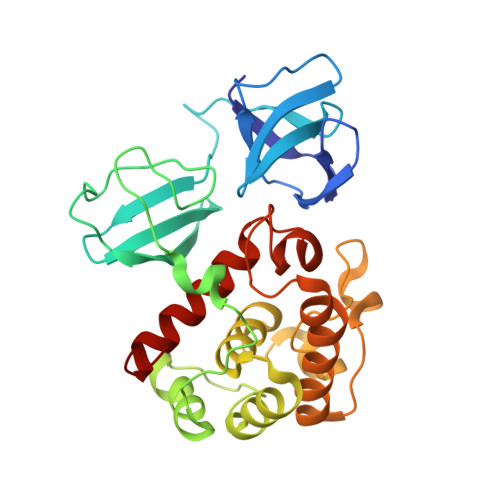
7PJ6

Crystal structure of catalytic domain of LytB (E585Q) from Streptococcus pneumoniae in complex with NAG-NAM-NAG-NAM-NAG peptidolycan analogue


Experimental Data Snapshot

  • Method: X-RAY DIFFRACTION
  • Resolution: 1.30 Å
  • R-Value Free: 0.174 
  • R-Value Work: 0.149 

Starting Model: experimental
View more details

wwPDB Validation   3D Report Full Report


This is version 1.3 of the entry. See complete history


Literature

Molecular basis of the final step of cell division in Streptococcus pneumoniae.

Martinez-Caballero, S.Freton, C.Molina, R.Bartual, S.G.Gueguen-Chaignon, V.Mercy, C.Gago, F.Mahasenan, K.V.Munoz, I.G.Lee, M.Hesek, D.Mobashery, S.Hermoso, J.A.Grangeasse, C.

(2023) Cell Rep 42: 112756-112756

  • DOI: https://doi.org/10.1016/j.celrep.2023.112756
  • Primary Citation of Related Structures:  
    7PJ3, 7PJ4, 7PJ5, 7PJ6, 7PL2, 7PL3, 7PL5, 7POD

  • PubMed Abstract: 

    Bacterial cell-wall hydrolases must be tightly regulated during bacterial cell division to prevent aberrant cell lysis and to allow final separation of viable daughter cells. In a multidisciplinary work, we disclose the molecular dialogue between the cell-wall hydrolase LytB, wall teichoic acids, and the eukaryotic-like protein kinase StkP in Streptococcus pneumoniae. After characterizing the peptidoglycan recognition mode by the catalytic domain of LytB, we further demonstrate that LytB possesses a modular organization allowing the specific binding to wall teichoic acids and to the protein kinase StkP. Structural and cellular studies notably reveal that the temporal and spatial localization of LytB is governed by the interaction between specific modules of LytB and the final PASTA domain of StkP. Our data collectively provide a comprehensive understanding of how LytB performs final separation of daughter cells and highlights the regulatory role of eukaryotic-like kinases on lytic machineries in the last step of cell division in streptococci.


  • Organizational Affiliation

    Department of Crystallography and Structural Biology, Instituto de Química-Física "Rocasolano," Consejo Superior de Investigaciones Científicas, Madrid, Spain.


Macromolecules
Find similar proteins by:  (by identity cutoff)  |  3D Structure
Entity ID: 1
MoleculeChains Sequence LengthOrganismDetailsImage
Putative endo-beta-N-acetylglucosaminidaseA [auth AAA]274Streptococcus pneumoniae R6Mutation(s): 1 
Gene Names: lytBspr0867
EC: 3.2.1.96
UniProt
Find proteins for P59206 (Streptococcus pneumoniae (strain ATCC BAA-255 / R6))
Explore P59206 
Go to UniProtKB:  P59206
Entity Groups  
Sequence Clusters30% Identity50% Identity70% Identity90% Identity95% Identity100% Identity
UniProt GroupP59206
Sequence Annotations
Expand
  • Reference Sequence
Oligosaccharides

Help

Entity ID: 2
MoleculeChains Length2D Diagram Glycosylation3D Interactions
2-acetamido-2-deoxy-beta-D-glucopyranose-(1-4)-N-acetyl-beta-muramic acid-(1-4)-2-acetamido-2-deoxy-beta-D-glucopyranose-(1-4)-N-acetyl-beta-muramic acid-(1-4)-2-acetamido-2-deoxy-beta-D-glucopyranoseB [auth AbA]5N/A
Experimental Data & Validation

Experimental Data

  • Method: X-RAY DIFFRACTION
  • Resolution: 1.30 Å
  • R-Value Free: 0.174 
  • R-Value Work: 0.149 
  • Space Group: C 2 2 21
Unit Cell:
Length ( Å )Angle ( ˚ )
a = 47.291α = 90
b = 92.543β = 90
c = 124.795γ = 90
Software Package:
Software NamePurpose
REFMACrefinement
XDSdata reduction
XDSdata scaling
PHASERphasing

Structure Validation

View Full Validation Report


Entry History & Funding Information

Deposition Data


Funding OrganizationLocationGrant Number
Not funded--

Revision History  (Full details and data files)

  • Version 1.0: 2022-09-07
    Type: Initial release
  • Version 1.1: 2023-07-12
    Changes: Database references, Structure summary
  • Version 1.2: 2023-07-19
    Changes: Database references
  • Version 1.3: 2024-02-07
    Changes: Data collection, Refinement description