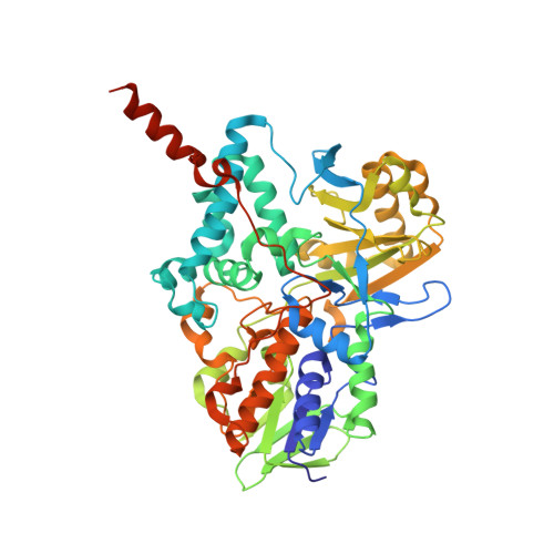
7P4F

Crystal Structure of Monoamine Oxidase B in complex with inhibitor 1


Experimental Data Snapshot

  • Method: X-RAY DIFFRACTION
  • Resolution: 2.30 Å
  • R-Value Free: 0.213 
  • R-Value Work: 0.161 
  • R-Value Observed: 0.163 

Starting Model: experimental
View more details

wwPDB Validation   3D Report Full Report


Ligand Structure Quality Assessment 


This is version 1.2 of the entry. See complete history


Literature

Dual Reversible Coumarin Inhibitors Mutually Bound to Monoamine Oxidase B and Acetylcholinesterase Crystal Structures.

Ekstrom, F.Gottinger, A.Forsgren, N.Catto, M.Iacovino, L.G.Pisani, L.Binda, C.

(2022) ACS Med Chem Lett 13: 499-506

  • DOI: https://doi.org/10.1021/acsmedchemlett.2c00001
  • Primary Citation of Related Structures:  
    7P4F, 7P4H, 7QAK, 7QB4

  • PubMed Abstract: 

    Multitarget directed ligands (MTDLs) represent a promising frontier in tackling the complexity of multifactorial pathologies. The synergistic inhibition of monoamine oxidase B (MAO B) and acetylcholinesterase (AChE) is believed to provide a potentiated effect in the treatment of Alzheimer's disease. Among previously reported micromolar or sub-micromolar coumarin-bearing dual inhibitors, compound 1 returned a tight-binding inhibition of MAO B ( K i = 4.5 μM) and a +5.5 °C increase in the enzyme T m value. Indeed, the X-ray crystal structure revealed that binding of 1 produces unforeseen conformational changes at the MAO B entrance cavity. Interestingly, 1 showed great shape complementarity with the AChE enzymatic gorge, being deeply buried from the catalytic anionic subsite (CAS) to the peripheral anionic subsite (PAS) and causing significant structural changes in the active site. These findings provide structural templates for further development of dual MAO B and AChE inhibitors.


  • Organizational Affiliation

    Swedish Defence Research Agency, CBRN Defence and Security, Umeå 901 82, Sweden.


Macromolecules
Find similar proteins by:  (by identity cutoff)  |  3D Structure
Entity ID: 1
MoleculeChains Sequence LengthOrganismDetailsImage
Amine oxidase [flavin-containing] B
A, B
520Homo sapiensMutation(s): 0 
Gene Names: MAOB
EC: 1.4.3.4 (PDB Primary Data), 1.4.3.21 (UniProt)
Membrane Entity: Yes 
UniProt & NIH Common Fund Data Resources
Find proteins for P27338 (Homo sapiens)
Explore P27338 
Go to UniProtKB:  P27338
PHAROS:  P27338
GTEx:  ENSG00000069535 
Entity Groups  
Sequence Clusters30% Identity50% Identity70% Identity90% Identity95% Identity100% Identity
UniProt GroupP27338
Sequence Annotations
Expand
  • Reference Sequence
Small Molecules
Ligands 3 Unique
IDChains Name / Formula / InChI Key2D Diagram3D Interactions
FAD
Query on FAD

Download Ideal Coordinates CCD File 
C [auth A],
F [auth B]
FLAVIN-ADENINE DINUCLEOTIDE
C27 H33 N9 O15 P2
VWWQXMAJTJZDQX-UYBVJOGSSA-N
5IK
Query on 5IK

Download Ideal Coordinates CCD File 
D [auth A],
G [auth B]
4-(hydroxymethyl)-7-[[4-[[methyl-(phenylmethyl)amino]methyl]phenyl]methoxy]chromen-2-one
C26 H25 N O4
RTJRBYKZQSYWCM-UHFFFAOYSA-N
C15
Query on C15

Download Ideal Coordinates CCD File 
E [auth A],
H [auth B]
N-DODECYL-N,N-DIMETHYL-3-AMMONIO-1-PROPANESULFONATE
C17 H38 N O3 S
IZWSFJTYBVKZNK-UHFFFAOYSA-O
Binding Affinity Annotations 
IDSourceBinding Affinity
5IK BindingDB:  7P4F IC50: 10 (nM) from 1 assay(s)
Experimental Data & Validation

Experimental Data

Unit Cell:
Length ( Å )Angle ( ˚ )
a = 131.243α = 90
b = 222.428β = 90
c = 86.206γ = 90
Software Package:
Software NamePurpose
REFMACrefinement
PDB_EXTRACTdata extraction
XDSdata reduction
Aimlessdata scaling
MOLREPphasing

Structure Validation

View Full Validation Report


Ligand Structure Quality Assessment 


Entry History 

Deposition Data

Revision History  (Full details and data files)

  • Version 1.0: 2022-05-18
    Type: Initial release
  • Version 1.1: 2024-01-31
    Changes: Data collection, Refinement description
  • Version 1.2: 2024-10-23
    Changes: Structure summary