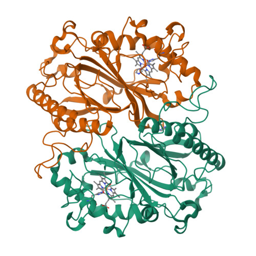
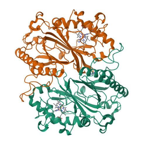
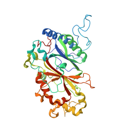
6I7C

Dye type peroxidase Aa from Streptomyces lividans: imidazole complex


Experimental Data Snapshot

  • Method: X-RAY DIFFRACTION
  • Resolution: 1.88 Å
  • R-Value Free: 0.177 
  • R-Value Work: 0.139 
  • R-Value Observed: 0.140 

Starting Model: experimental
View more details

wwPDB Validation   3D Report Full Report


Ligand Structure Quality Assessment 

Created with Raphaël 2.3.0Worse 01 BetterLigand structure goodness of fit to experimental dataBest fitted HEMClick on this verticalbar to view details

This is version 1.2 of the entry. See complete history


Literature

High-throughput structures of protein-ligand complexes at room temperature using serial femtosecond crystallography.

Moreno-Chicano, T.Ebrahim, A.Axford, D.Appleby, M.V.Beale, J.H.Chaplin, A.K.Duyvesteyn, H.M.E.Ghiladi, R.A.Owada, S.Sherrell, D.A.Strange, R.W.Sugimoto, H.Tono, K.Worrall, J.A.R.Owen, R.L.Hough, M.A.

(2019) IUCrJ 6: 1074-1085

  • DOI: https://doi.org/10.1107/S2052252519011655
  • Primary Citation of Related Structures:  
    6I6G, 6I7C, 6I7F, 6QWG

  • PubMed Abstract: 

    High-throughput X-ray crystal structures of protein-ligand complexes are critical to pharmaceutical drug development. However, cryocooling of crystals and X-ray radiation damage may distort the observed ligand binding. Serial femtosecond crystallography (SFX) using X-ray free-electron lasers (XFELs) can produce radiation-damage-free room-temperature structures. Ligand-binding studies using SFX have received only modest attention, partly owing to limited beamtime availability and the large quantity of sample that is required per structure determination. Here, a high-throughput approach to determine room-temperature damage-free structures with excellent sample and time efficiency is demonstrated, allowing complexes to be characterized rapidly and without prohibitive sample requirements. This yields high-quality difference density maps allowing unambiguous ligand placement. Crucially, it is demonstrated that ligands similar in size or smaller than those used in fragment-based drug design may be clearly identified in data sets obtained from <1000 diffraction images. This efficiency in both sample and XFEL beamtime opens the door to true high-throughput screening of protein-ligand complexes using SFX.


  • Organizational Affiliation

    School of Life Sciences, University of Essex, Wivenhoe Park, Colchester CO4 3SQ, England.


Macromolecules
Find similar proteins by:  (by identity cutoff)  |  3D Structure
Entity ID: 1
MoleculeChains Sequence LengthOrganismDetailsImage
Deferrochelatase/peroxidase
A, B
363Streptomyces coelicolor A3(2)Mutation(s): 0 
Gene Names: SCO2276
EC: 1.11.1
UniProt
Find proteins for Q9RKQ2 (Streptomyces coelicolor (strain ATCC BAA-471 / A3(2) / M145))
Explore Q9RKQ2 
Go to UniProtKB:  Q9RKQ2
Entity Groups  
Sequence Clusters30% Identity50% Identity70% Identity90% Identity95% Identity100% Identity
UniProt GroupQ9RKQ2
Sequence Annotations
Expand
  • Reference Sequence
Experimental Data & Validation

Experimental Data

  • Method: X-RAY DIFFRACTION
  • Resolution: 1.88 Å
  • R-Value Free: 0.177 
  • R-Value Work: 0.139 
  • R-Value Observed: 0.140 
  • Space Group: P 1 21 1
Unit Cell:
Length ( Å )Angle ( ˚ )
a = 72.48α = 90
b = 68.03β = 105.57
c = 73.53γ = 90
Software Package:
Software NamePurpose
PHENIXrefinement
PDB_EXTRACTdata extraction
CrystFELdata reduction
CrystFELdata scaling
MOLREPphasing

Structure Validation

View Full Validation Report


Ligand Structure Quality Assessment 

Created with Raphaël 2.3.0Worse 01 BetterLigand structure goodness of fit to experimental dataBest fitted HEMClick on this verticalbar to view details

Entry History 

Deposition Data

Revision History  (Full details and data files)

  • Version 1.0: 2019-11-20
    Type: Initial release
  • Version 1.1: 2020-01-22
    Changes: Data collection
  • Version 1.2: 2024-01-24
    Changes: Data collection, Database references, Derived calculations, Refinement description