6G2S | pdb_00006g2s

Crystal structure of FimH in complex with a pentaflourinated biphenyl alpha D-mannoside


Experimental Data Snapshot

  • Method: X-RAY DIFFRACTION
  • Resolution: 2.20 Å
  • R-Value Free: 
    0.222 (Depositor), 0.220 (DCC) 
  • R-Value Work: 
    0.197 (Depositor), 0.200 (DCC) 
  • R-Value Observed: 
    0.198 (Depositor) 

Starting Model: experimental
View more details

wwPDB Validation   3D Report Full Report


Ligand Structure Quality Assessment 

Created with Raphaël 2.3.0Worse 01 BetterLigand structure goodness of fit to experimental dataBest fitted EJNClick on this verticalbar to view details

This is version 1.3 of the entry. See complete history


Literature

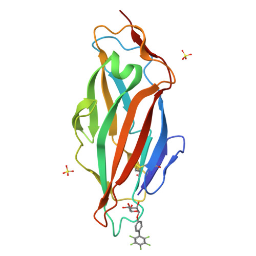
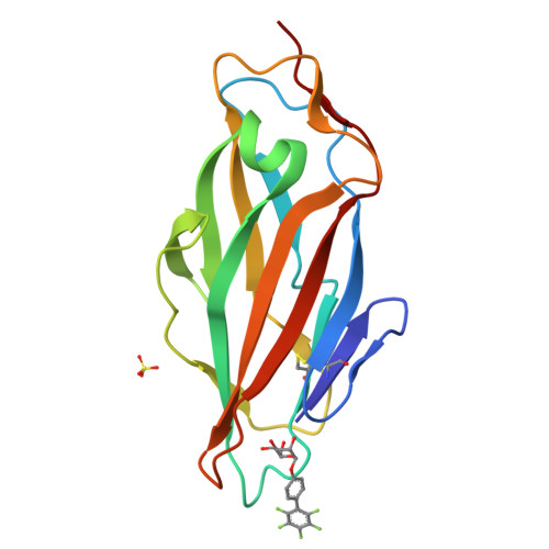
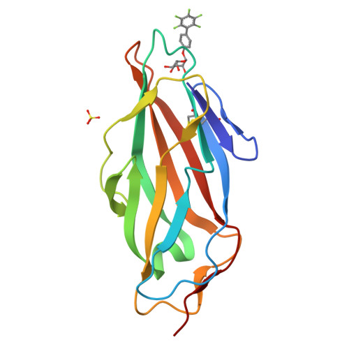
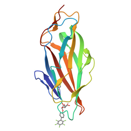
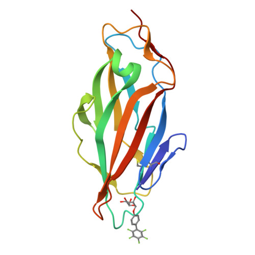
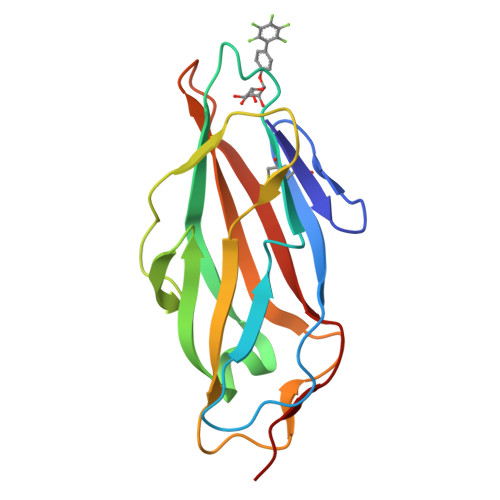
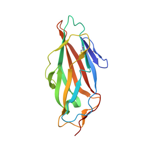
Improvement of Aglycone pi-Stacking Yields Nanomolar to Sub-nanomolar FimH Antagonists.

Schonemann, W.Cramer, J.Muhlethaler, T.Fiege, B.Silbermann, M.Rabbani, S.Datwyler, P.Zihlmann, P.Jakob, R.P.Sager, C.P.Smiesko, M.Schwardt, O.Maier, T.Ernst, B.

(2019) ChemMedChem 14: 749-757

  • DOI: https://doi.org/10.1002/cmdc.201900051
  • Primary Citation of Related Structures:  
    6G2R, 6G2S

  • PubMed Abstract: 

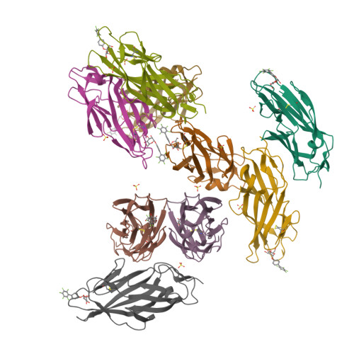
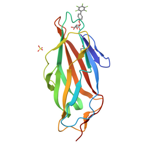
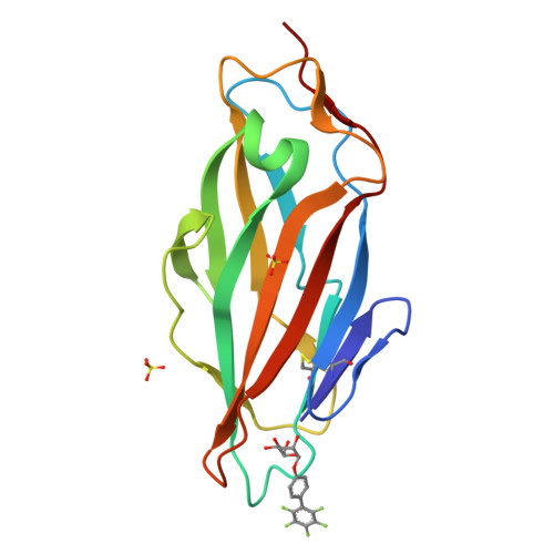
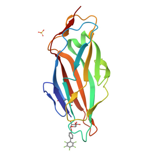
    Antimicrobial resistance has become a serious concern for the treatment of urinary tract infections. In this context, an anti-adhesive approach targeting FimH, a bacterial lectin enabling the attachment of E. coli to host cells, has attracted considerable interest. FimH can adopt a low/medium-affinity state in the absence and a high-affinity state in the presence of shear forces. Until recently, mostly the high-affinity state has been investigated, despite the fact that a therapeutic antagonist should bind predominantly to the low-affinity state. In this communication, we demonstrate that fluorination of biphenyl α-d-mannosides leads to compounds with perfect π-π stacking interactions with the tyrosine gate of FimH, yielding low nanomolar to sub-nanomolar K D values for the low- and high-affinity states, respectively. The face-to-face alignment of the perfluorinated biphenyl group of FimH ligands and Tyr48 was confirmed by crystal structures as well as 1 H, 15 N-HSQC NMR analysis. Finally, fluorination improves pharmacokinetic parameters predictive for oral availability.


  • Organizational Affiliation

    Department of Pharmaceutical Sciences, University of Basel, Klingelbergstrasse 50, 4056, Basel, Switzerland.


Macromolecules
Find similar proteins by:  (by identity cutoff)  |  3D Structure
Entity ID: 1
MoleculeChains Sequence LengthOrganismDetailsImage
Type 1 fimbrin D-mannose specific adhesin
A, B, C, D, E
158Escherichia coli K-12Mutation(s): 0 
Gene Names: fimHb4320JW4283
UniProt
Find proteins for P08191 (Escherichia coli (strain K12))
Explore P08191 
Go to UniProtKB:  P08191
Entity Groups  
Sequence Clusters30% Identity50% Identity70% Identity90% Identity95% Identity100% Identity
UniProt GroupP08191
Sequence Annotations
Expand
  • Reference Sequence
Small Molecules
Ligands 2 Unique
IDChains Name / Formula / InChI Key2D Diagram3D Interactions
EJN
Query on EJN

Download Ideal Coordinates CCD File 
AA [auth I]
J [auth A]
L [auth B]
O [auth C]
Q [auth D]
(2~{R},3~{S},4~{S},5~{S},6~{R})-2-(hydroxymethyl)-6-[4-[2,3,4,5,6-pentakis(fluoranyl)phenyl]phenoxy]oxane-3,4,5-triol
C18 H15 F5 O6
WCSHODXKNWBNJL-VYMSMKAZSA-N
SO4
Query on SO4

Download Ideal Coordinates CCD File 
K [auth A]
M [auth B]
N [auth B]
P [auth C]
R [auth D]
SULFATE ION
O4 S
QAOWNCQODCNURD-UHFFFAOYSA-L
Experimental Data & Validation

Experimental Data

  • Method: X-RAY DIFFRACTION
  • Resolution: 2.20 Å
  • R-Value Free:  0.222 (Depositor), 0.220 (DCC) 
  • R-Value Work:  0.197 (Depositor), 0.200 (DCC) 
  • R-Value Observed: 0.198 (Depositor) 
Space Group: P 32
Unit Cell:
Length ( Å )Angle ( ˚ )
a = 86.01α = 90
b = 86.01β = 90
c = 203.14γ = 120
Software Package:
Software NamePurpose
PHENIXrefinement
XDSdata reduction
XSCALEdata scaling
PHASERphasing

Structure Validation

View Full Validation Report


Ligand Structure Quality Assessment 

Created with Raphaël 2.3.0Worse 01 BetterLigand structure goodness of fit to experimental dataBest fitted EJNClick on this verticalbar to view details

Entry History 

Revision History  (Full details and data files)

  • Version 1.0: 2019-03-20
    Type: Initial release
  • Version 1.1: 2019-04-17
    Changes: Data collection, Database references
  • Version 1.2: 2024-01-17
    Changes: Data collection, Database references, Refinement description
  • Version 1.3: 2024-11-13
    Changes: Structure summary