5VYY | pdb_00005vyy

Structure of human Hsp90-alpha bound to resorcinylic inhibitor BnIm


Experimental Data Snapshot

  • Method: X-RAY DIFFRACTION
  • Resolution: 1.79 Å
  • R-Value Free: 
    0.218 (Depositor), 0.220 (DCC) 
  • R-Value Work: 
    0.184 (Depositor), 0.180 (DCC) 
  • R-Value Observed: 
    0.187 (Depositor) 

wwPDB Validation   3D Report Full Report


Ligand Structure Quality Assessment 

Created with Raphaël 2.3.0Worse 01 BetterLigand structure goodness of fit to experimental dataBest fitted 9QYClick on this verticalbar to view details

This is version 1.3 of the entry. See complete history


Literature

Structure Based Design of a Grp94-Selective Inhibitor: Exploiting a Key Residue in Grp94 To Optimize Paralog-Selective Binding.

Que, N.L.S.Crowley, V.M.Duerfeldt, A.S.Zhao, J.Kent, C.N.Blagg, B.S.J.Gewirth, D.T.

(2018) J Med Chem 61: 2793-2805

  • DOI: https://doi.org/10.1021/acs.jmedchem.7b01608
  • Primary Citation of Related Structures:  
    5VYY, 5WMT, 6BAW, 6C91, 6CEO

  • PubMed Abstract: 

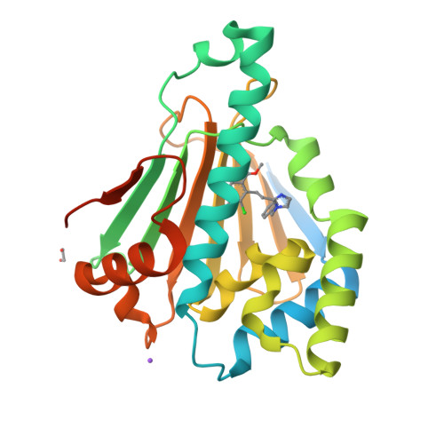
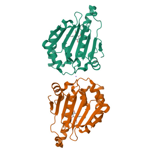
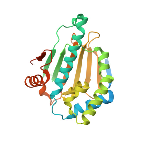
    Grp94 and Hsp90, the ER and cytoplasmic hsp90 paralogs, share a conserved ATP-binding pocket that has been targeted for therapeutics. Paralog-selective inhibitors may lead to drugs with fewer side effects. Here, we analyzed 1 (BnIm), a benzyl imidazole resorcinylic inhibitor, for its mode of binding. The structures of 1 bound to Hsp90 and Grp94 reveal large conformational changes in Grp94 but not Hsp90 that expose site 2, a binding pocket adjacent to the central ATP cavity that is ordinarily blocked. The Grp94:1 structure reveals a flipped pose of the resorcinylic scaffold that inserts into the exposed site 2. We exploited this flipped binding pose to develop a Grp94-selective derivative of 1. Our structural analysis shows that the ability of the ligand to insert its benzyl imidazole substituent into site 1, a different side pocket off the ATP binding cavity, is the key to exposing site 2 in Grp94.


  • Organizational Affiliation

    Hauptman-Woodward Medical Research Institute , Buffalo , New York 14203 , United States.


Macromolecules
Find similar proteins by:  (by identity cutoff)  |  3D Structure
Entity ID: 1
MoleculeChains Sequence LengthOrganismDetailsImage
Heat shock protein HSP 90-alpha256Homo sapiensMutation(s): 0 
Gene Names: HSP90AA1HSP90AHSPC1HSPCA
EC: 3.6.4.10
UniProt & NIH Common Fund Data Resources
Find proteins for P07900 (Homo sapiens)
Explore P07900 
Go to UniProtKB:  P07900
PHAROS:  P07900
GTEx:  ENSG00000080824 
Entity Groups  
Sequence Clusters30% Identity50% Identity70% Identity90% Identity95% Identity100% Identity
UniProt GroupP07900
Sequence Annotations
Expand
  • Reference Sequence
Small Molecules
Ligands 5 Unique
IDChains Name / Formula / InChI Key2D Diagram3D Interactions
9QY
Query on 9QY

Download Ideal Coordinates CCD File 
B [auth A]methyl 2-[2-(1-benzyl-1H-imidazol-2-yl)ethyl]-3-chloro-4,6-dihydroxybenzoate
C20 H19 Cl N2 O4
NCWIQXPFJORNJP-UHFFFAOYSA-N
EDO
Query on EDO

Download Ideal Coordinates CCD File 
E [auth A]1,2-ETHANEDIOL
C2 H6 O2
LYCAIKOWRPUZTN-UHFFFAOYSA-N
FMT
Query on FMT

Download Ideal Coordinates CCD File 
C [auth A],
D [auth A]
FORMIC ACID
C H2 O2
BDAGIHXWWSANSR-UHFFFAOYSA-N
MG
Query on MG

Download Ideal Coordinates CCD File 
F [auth A]MAGNESIUM ION
Mg
JLVVSXFLKOJNIY-UHFFFAOYSA-N
NA
Query on NA

Download Ideal Coordinates CCD File 
G [auth A]SODIUM ION
Na
FKNQFGJONOIPTF-UHFFFAOYSA-N
Binding Affinity Annotations 
IDSourceBinding Affinity
9QY BindingDB:  5VYY Ki: 1800 (nM) from 1 assay(s)
Kd: min: 620, max: 1.31e+4 (nM) from 2 assay(s)
IC50: min: 1.31e+4, max: 1.40e+4 (nM) from 3 assay(s)
Experimental Data & Validation

Experimental Data

  • Method: X-RAY DIFFRACTION
  • Resolution: 1.79 Å
  • R-Value Free:  0.218 (Depositor), 0.220 (DCC) 
  • R-Value Work:  0.184 (Depositor), 0.180 (DCC) 
  • R-Value Observed: 0.187 (Depositor) 
Space Group: C 2 2 21
Unit Cell:
Length ( Å )Angle ( ˚ )
a = 67.075α = 90
b = 92.055β = 90
c = 85.262γ = 90
Software Package:
Software NamePurpose
PHENIXrefinement
HKL-2000data reduction
HKL-2000data scaling
PHENIXphasing

Structure Validation

View Full Validation Report


Ligand Structure Quality Assessment 

Created with Raphaël 2.3.0Worse 01 BetterLigand structure goodness of fit to experimental dataBest fitted 9QYClick on this verticalbar to view details

Entry History & Funding Information

Deposition Data


Funding OrganizationLocationGrant Number
National Institutes of Health/National Cancer Institute (NIH/NCI)United StatesR01-CA095130
National Institutes of Health/National Cancer Institute (NIH/NCI)United StatesP01-CA186866

Revision History  (Full details and data files)

  • Version 1.0: 2018-04-18
    Type: Initial release
  • Version 1.1: 2018-04-25
    Changes: Data collection, Database references
  • Version 1.2: 2019-12-04
    Changes: Author supporting evidence
  • Version 1.3: 2024-03-13
    Changes: Data collection, Database references, Derived calculations