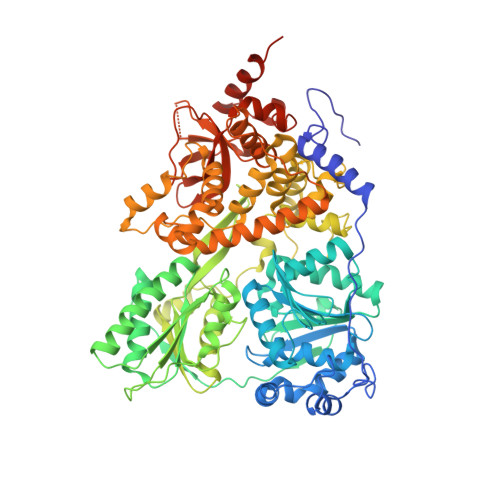
5JPT

CRYSTAL STRUCTURE OF THE PRP43P DEAH-BOX RNA HELICASE IN COMPLEX WITH CDP


Experimental Data Snapshot

  • Method: X-RAY DIFFRACTION
  • Resolution: 2.94 Å
  • R-Value Free: 0.257 
  • R-Value Work: 0.203 
  • R-Value Observed: 0.206 

Starting Model: experimental
View more details

wwPDB Validation   3D Report Full Report


Ligand Structure Quality Assessment 


This is version 1.1 of the entry. See complete history


Literature

Functional link between DEAH/RHA helicase Prp43 activation and ATP base binding.

Robert-Paganin, J.Halladjian, M.Blaud, M.Lebaron, S.Delbos, L.Chardon, F.Capeyrou, R.Humbert, O.Henry, Y.Henras, A.K.Rety, S.Leulliot, N.

(2017) Nucleic Acids Res 45: 1539-1552

  • DOI: https://doi.org/10.1093/nar/gkw1233
  • Primary Citation of Related Structures:  
    5JPT

  • PubMed Abstract: 

    The DEAH box helicase Prp43 is a bifunctional enzyme from the DEAH/RHA helicase family required both for the maturation of ribosomes and for lariat intron release during splicing. It interacts with G-patch domain containing proteins which activate the enzymatic activity of Prp43 in vitro by an unknown mechanism. In this work, we show that the activation by G-patch domains is linked to the unique nucleotide binding mode of this helicase family. The base of the ATP molecule is stacked between two residues, R159 of the RecA1 domain (R-motif) and F357 of the RecA2 domain (F-motif). Using Prp43 F357A mutants or pyrimidine nucleotides, we show that the lack of stacking of the nucleotide base to the F-motif decouples the NTPase and helicase activities of Prp43. In contrast the R159A mutant (R-motif) showed reduced ATPase and helicase activities. We show that the Prp43 R-motif mutant induces the same phenotype as the absence of the G-patch protein Gno1, strongly suggesting that the processing defects observed in the absence of Gno1 result from a failure to activate the Prp43 helicase. Overall we propose that the stacking between the R- and F-motifs and the nucleotide base is important for the activity and regulation of this helicase family.


  • Organizational Affiliation

    Laboratoire de Cristallographie et RMN Biologiques, UMR CNRS 8015, Université Paris Descartes, Sorbonne Paris Cité, Faculté de Pharmacie, Paris, France.


Macromolecules
Find similar proteins by:  (by identity cutoff)  |  3D Structure
Entity ID: 1
MoleculeChains Sequence LengthOrganismDetailsImage
Pre-mRNA-splicing factor ATP-dependent RNA helicase PRP43
A, B
767Saccharomyces cerevisiae S288CMutation(s): 0 
Gene Names: PRP43YGL120C
EC: 3.6.4.13
UniProt
Find proteins for P53131 (Saccharomyces cerevisiae (strain ATCC 204508 / S288c))
Explore P53131 
Go to UniProtKB:  P53131
Entity Groups  
Sequence Clusters30% Identity50% Identity70% Identity90% Identity95% Identity100% Identity
UniProt GroupP53131
Sequence Annotations
Expand
  • Reference Sequence
Small Molecules
Ligands 5 Unique
IDChains Name / Formula / InChI Key2D Diagram3D Interactions
CDP
Query on CDP

Download Ideal Coordinates CCD File 
C [auth A],
H [auth B]
CYTIDINE-5'-DIPHOSPHATE
C9 H15 N3 O11 P2
ZWIADYZPOWUWEW-XVFCMESISA-N
GOL
Query on GOL

Download Ideal Coordinates CCD File 
E [auth A]GLYCEROL
C3 H8 O3
PEDCQBHIVMGVHV-UHFFFAOYSA-N
ACT
Query on ACT

Download Ideal Coordinates CCD File 
F [auth A]ACETATE ION
C2 H3 O2
QTBSBXVTEAMEQO-UHFFFAOYSA-M
NI
Query on NI

Download Ideal Coordinates CCD File 
G [auth A]NICKEL (II) ION
Ni
VEQPNABPJHWNSG-UHFFFAOYSA-N
MG
Query on MG

Download Ideal Coordinates CCD File 
D [auth A],
I [auth B]
MAGNESIUM ION
Mg
JLVVSXFLKOJNIY-UHFFFAOYSA-N
Experimental Data & Validation

Experimental Data

  • Method: X-RAY DIFFRACTION
  • Resolution: 2.94 Å
  • R-Value Free: 0.257 
  • R-Value Work: 0.203 
  • R-Value Observed: 0.206 
  • Space Group: P 32 2 1
Unit Cell:
Length ( Å )Angle ( ˚ )
a = 118.07α = 90
b = 118.07β = 90
c = 252.15γ = 120
Software Package:
Software NamePurpose
PHENIXrefinement
XDSdata reduction
XDSdata scaling
PHASERphasing

Structure Validation

View Full Validation Report


Ligand Structure Quality Assessment 


Entry History 

Deposition Data

Revision History  (Full details and data files)

  • Version 1.0: 2017-02-22
    Type: Initial release
  • Version 1.1: 2024-01-10
    Changes: Data collection, Database references, Derived calculations, Refinement description