5IJC

The crystal structure of mouse TLR4/MD-2/neoseptin-3 complex


Experimental Data Snapshot

  • Method: X-RAY DIFFRACTION
  • Resolution: 2.57 Å
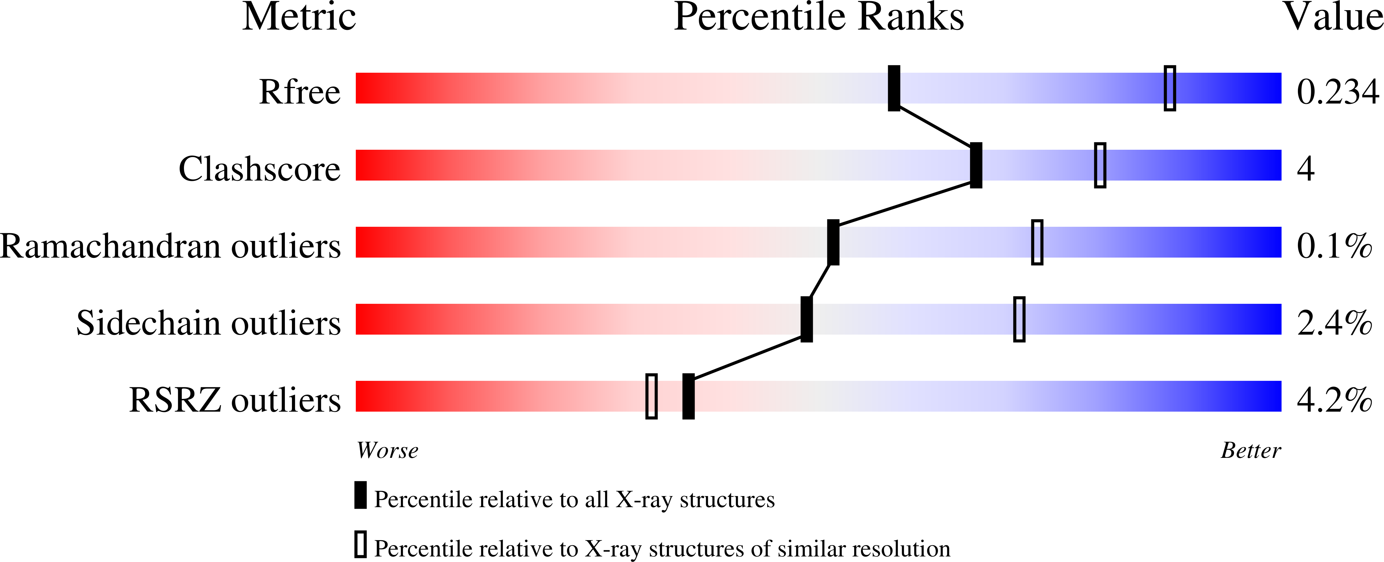
  • R-Value Free: 0.234 
  • R-Value Work: 0.193 
  • R-Value Observed: 0.195 

Starting Model: experimental
View more details

wwPDB Validation   3D Report Full Report


Ligand Structure Quality Assessment 


This is version 3.2 of the entry. See complete history


Literature

TLR4/MD-2 activation by a synthetic agonist with no similarity to LPS.

Wang, Y.Su, L.Morin, M.D.Jones, B.T.Whitby, L.R.Surakattula, M.M.Huang, H.Shi, H.Choi, J.H.Wang, K.W.Moresco, E.M.Berger, M.Zhan, X.Zhang, H.Boger, D.L.Beutler, B.

(2016) Proc Natl Acad Sci U S A 113: E884-E893

  • DOI: https://doi.org/10.1073/pnas.1525639113
  • Primary Citation of Related Structures:  
    5IJB, 5IJC, 5IJD

  • PubMed Abstract: 

    Structurally disparate molecules reportedly engage and activate Toll-like receptor (TLR) 4 and other TLRs, yet the interactions that mediate binding and activation by dissimilar ligands remain unknown. We describe Neoseptins, chemically synthesized peptidomimetics that bear no structural similarity to the established TLR4 ligand, lipopolysaccharide (LPS), but productively engage the mouse TLR4 (mTLR4)/myeloid differentiation factor 2 (MD-2) complex. Neoseptin-3 activates mTLR4/MD-2 independently of CD14 and triggers canonical myeloid differentiation primary response gene 88 (MyD88)- and Toll-interleukin 1 receptor (TIR) domain-containing adaptor inducing IFN-beta (TRIF)-dependent signaling. The crystal structure mTLR4/MD-2/Neoseptin-3 at 2.57-Å resolution reveals that Neoseptin-3 binds as an asymmetrical dimer within the hydrophobic pocket of MD-2, inducing an active receptor complex similar to that induced by lipid A. However, Neoseptin-3 and lipid A form dissimilar molecular contacts to achieve receptor activation; hence strong TLR4/MD-2 agonists need not mimic LPS.


  • Organizational Affiliation

    Center for the Genetics of Host Defense, University of Texas Southwestern Medical Center, Dallas, TX 75390;


Macromolecules
Find similar proteins by:  (by identity cutoff)  |  3D Structure
Entity ID: 1
MoleculeChains Sequence LengthOrganismDetailsImage
Toll-like receptor 4,Variable lymphocyte receptor BA,
C [auth B]
635Mus musculusEptatretus burgeri
This entity is chimeric
Mutation(s): 0 
Gene Names: Tlr4LpsVLRB
EC: 3.2.2.6
UniProt & NIH Common Fund Data Resources
Find proteins for Q4G1L2 (Eptatretus burgeri)
Explore Q4G1L2 
Go to UniProtKB:  Q4G1L2
Find proteins for Q9QUK6 (Mus musculus)
Explore Q9QUK6 
Go to UniProtKB:  Q9QUK6
IMPC:  MGI:96824
Entity Groups  
Sequence Clusters30% Identity50% Identity70% Identity90% Identity95% Identity100% Identity
UniProt GroupsQ4G1L2Q9QUK6
Glycosylation
Glycosylation Sites: 6Go to GlyGen: Q9QUK6-1
Sequence Annotations
Expand
  • Reference Sequence
Find similar proteins by:  (by identity cutoff)  |  3D Structure
Entity ID: 2
MoleculeChains Sequence LengthOrganismDetailsImage
Lymphocyte antigen 96B [auth C],
D
191Mus musculusMutation(s): 0 
Gene Names: Ly96Esop1Md2
UniProt
Find proteins for Q9JHF9 (Mus musculus)
Explore Q9JHF9 
Go to UniProtKB:  Q9JHF9
Entity Groups  
Sequence Clusters30% Identity50% Identity70% Identity90% Identity95% Identity100% Identity
UniProt GroupQ9JHF9
Glycosylation
Glycosylation Sites: 2Go to GlyGen: Q9JHF9-1
Sequence Annotations
Expand
  • Reference Sequence
Oligosaccharides

Help

Entity ID: 3
MoleculeChains Length2D Diagram Glycosylation3D Interactions
2-acetamido-2-deoxy-beta-D-glucopyranose-(1-4)-2-acetamido-2-deoxy-beta-D-glucopyranose
E, F, G, H, I
2N-Glycosylation
Glycosylation Resources
GlyTouCan:  G42666HT
GlyCosmos:  G42666HT
GlyGen:  G42666HT
Small Molecules
Ligands 2 Unique
IDChains Name / Formula / InChI Key2D Diagram3D Interactions
V2A (Subject of Investigation/LOI)
Query on V2A

Download Ideal Coordinates CCD File 
N [auth A],
P [auth B],
Q [auth B],
V [auth D]
neoseptin 3
C29 H34 N2 O4
OACODUCFPHHCIH-SANMLTNESA-N
NAG
Query on NAG

Download Ideal Coordinates CCD File 
J [auth A]
K [auth A]
L [auth A]
M [auth A]
O [auth C]
J [auth A],
K [auth A],
L [auth A],
M [auth A],
O [auth C],
R [auth B],
S [auth B],
T [auth B],
U [auth B],
W [auth D],
X [auth D]
2-acetamido-2-deoxy-beta-D-glucopyranose
C8 H15 N O6
OVRNDRQMDRJTHS-FMDGEEDCSA-N
Binding Affinity Annotations 
IDSourceBinding Affinity
V2A BindingDB:  5IJC IC50: 1.85e+4 (nM) from 1 assay(s)
Experimental Data & Validation

Experimental Data

  • Method: X-RAY DIFFRACTION
  • Resolution: 2.57 Å
  • R-Value Free: 0.234 
  • R-Value Work: 0.193 
  • R-Value Observed: 0.195 
  • Space Group: P 1 21 1
Unit Cell:
Length ( Å )Angle ( ˚ )
a = 70.752α = 90
b = 148.559β = 98.304
c = 131.72γ = 90
Software Package:
Software NamePurpose
PHENIXrefinement
PHENIXrefinement
HKL-3000data reduction
HKL-3000data scaling
PHASERphasing

Structure Validation

View Full Validation Report


Ligand Structure Quality Assessment 


Entry History 

Deposition Data

Revision History  (Full details and data files)

  • Version 1.0: 2016-04-27
    Type: Initial release
  • Version 1.1: 2016-05-04
    Changes: Database references
  • Version 2.0: 2020-07-01
    Changes: Advisory, Atomic model, Author supporting evidence, Data collection, Database references, Derived calculations, Experimental preparation, Non-polymer description, Other, Polymer sequence, Refinement description, Source and taxonomy, Structure summary
  • Version 3.0: 2020-07-29
    Type: Remediation
    Reason: Carbohydrate remediation
    Changes: Atomic model, Data collection, Derived calculations, Structure summary
  • Version 3.1: 2023-09-27
    Changes: Data collection, Database references, Refinement description, Structure summary
  • Version 3.2: 2024-11-13
    Changes: Structure summary