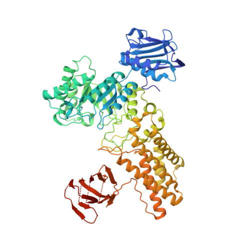
5FL0

Structure of a hydrolase with an inhibitor


Experimental Data Snapshot

  • Method: X-RAY DIFFRACTION
  • Resolution: 1.95 Å
  • R-Value Free: 0.227 
  • R-Value Work: 0.200 
  • R-Value Observed: 0.201 

wwPDB Validation   3D Report Full Report


Ligand Structure Quality Assessment 


This is version 2.0 of the entry. See complete history


Literature

Analysis of transition state mimicry by tight binding aminothiazoline inhibitors provides insight into catalysis by humanO-GlcNAcase.

Cekic, N.Heinonen, J.E.Stubbs, K.A.Roth, C.He, Y.Bennet, A.J.McEachern, E.J.Davies, G.J.Vocadlo, D.J.

(2016) Chem Sci 7: 3742-3750

  • DOI: https://doi.org/10.1039/c6sc00370b
  • Primary Citation of Related Structures:  
    5FKY, 5FL0, 5FL1

  • PubMed Abstract: 

    The modification of nucleocytoplasmic proteins with O -linked N -acetylglucosamine ( O -GlcNAc) plays diverse roles in multicellular organisms. Inhibitors of O -GlcNAc hydrolase (OGA), the enzyme that removes O -GlcNAc from proteins, lead to increased O -GlcNAc levels in cells and are seeing widespread adoption in the field as a research tool used in cells and in vivo . Here we synthesize and study a series of tight binding carbohydrate-based inhibitors of human OGA (hOGA). The most potent of these 2'-aminothiazolines binds with a sub-nanomolar K i value to hOGA (510 ± 50 pM) and the most selective has greater than 1 800 000-fold selectivity for hOGA over mechanistically related human lysosomal β-hexosaminidase. Structural data of inhibitors in complex with an hOGA homologue reveals the basis for variation in binding among these compounds. Using linear free energy analyses, we show binding of these 2'-aminothiazoline inhibitors depends on the p K a of the aminothiazoline ring system, revealing the protonation state of the inhibitor is a key driver of binding. Using series of inhibitors and synthetic substrates, we show that 2'-aminothiazoline inhibitors are transition state analogues of hOGA that bind to the enzyme up to 1-million fold more tightly than the substrate. These collective data support an oxazoline, rather than a protonated oxazolinium ion, intermediate being formed along the reaction pathway. Inhibitors from this series will prove generally useful tools for the study of O -GlcNAc. The new insights gained here, into the catalytic mechanism of hOGA and the fundamental drivers of potency and selectivity of OGA inhibitors, should enable tuning of hOGA inhibitors with desirable properties.


  • Organizational Affiliation

    Department of Chemistry , Simon Fraser University , Burnaby , British Columbia V5A 1S6 , Canada . Email: [email protected].


Macromolecules
Find similar proteins by:  (by identity cutoff)  |  3D Structure
Entity ID: 1
MoleculeChains Sequence LengthOrganismDetailsImage
O-GLCNACASE BT_4395
A, B
716Bacteroides thetaiotaomicronMutation(s): 0 
EC: 3.2.1.169
UniProt
Find proteins for Q89ZI2 (Bacteroides thetaiotaomicron (strain ATCC 29148 / DSM 2079 / JCM 5827 / CCUG 10774 / NCTC 10582 / VPI-5482 / E50))
Explore Q89ZI2 
Go to UniProtKB:  Q89ZI2
Entity Groups  
Sequence Clusters30% Identity50% Identity70% Identity90% Identity95% Identity100% Identity
UniProt GroupQ89ZI2
Sequence Annotations
Expand
  • Reference Sequence
Small Molecules
Ligands 3 Unique
IDChains Name / Formula / InChI Key2D Diagram3D Interactions
71I
Query on 71I

Download Ideal Coordinates CCD File 
E [auth A],
I [auth B]
(3~{a}~{R},5~{R},6~{S},7~{R},7~{a}~{R})-2-(butylamino)-5-(hydroxymethyl)-5,6,7,7~{a}-tetrahydro-3~{a}~{H}-pyrano[3,2-d] [1,3]thiazole-6,7-diol
C11 H20 N2 O4 S
KFNMCGAJIQGULW-VVULQXIFSA-N
EDO
Query on EDO

Download Ideal Coordinates CCD File 
C [auth A],
D [auth A],
G [auth B],
H [auth B]
1,2-ETHANEDIOL
C2 H6 O2
LYCAIKOWRPUZTN-UHFFFAOYSA-N
CA
Query on CA

Download Ideal Coordinates CCD File 
F [auth B]CALCIUM ION
Ca
BHPQYMZQTOCNFJ-UHFFFAOYSA-N
Experimental Data & Validation

Experimental Data

  • Method: X-RAY DIFFRACTION
  • Resolution: 1.95 Å
  • R-Value Free: 0.227 
  • R-Value Work: 0.200 
  • R-Value Observed: 0.201 
  • Space Group: P 2 21 21
Unit Cell:
Length ( Å )Angle ( ˚ )
a = 51.529α = 90
b = 162.129β = 90
c = 223.643γ = 90
Software Package:
Software NamePurpose
PHENIXrefinement
XDSdata reduction
XDSdata scaling

Structure Validation

View Full Validation Report


Ligand Structure Quality Assessment 


Entry History 

Deposition Data

Revision History  (Full details and data files)

  • Version 1.0: 2016-08-31
    Type: Initial release
  • Version 1.1: 2018-08-29
    Changes: Data collection, Database references
  • Version 2.0: 2024-05-08
    Changes: Atomic model, Data collection, Database references, Derived calculations, Other, Structure summary