5EGS

Human PRMT6 with bound fragment-type inhibitor


Experimental Data Snapshot

  • Method: X-RAY DIFFRACTION
  • Resolution: 2.15 Å
  • R-Value Free: 
    0.246 (Depositor), 0.250 (DCC) 
  • R-Value Work: 
    0.201 (Depositor), 0.210 (DCC) 
  • R-Value Observed: 
    0.202 (Depositor) 

Starting Model: experimental
View more details

wwPDB Validation   3D Report Full Report


Ligand Structure Quality Assessment 

Created with Raphaël 2.3.0Worse 01 BetterLigand structure goodness of fit to experimental dataBest fitted SAHClick on this verticalbar to view detailsBest fitted 5NRClick on this verticalbar to view details

This is version 1.2 of the entry. See complete history


Literature

Discovery of a Potent Class I Protein Arginine Methyltransferase Fragment Inhibitor.

Ferreira de Freitas, R.Eram, M.S.Szewczyk, M.M.Steuber, H.Smil, D.Wu, H.Li, F.Senisterra, G.Dong, A.Brown, P.J.Hitchcock, M.Moosmayer, D.Stegmann, C.M.Egner, U.Arrowsmith, C.Barsyte-Lovejoy, D.Vedadi, M.Schapira, M.

(2016) J Med Chem 59: 1176-1183

  • DOI: https://doi.org/10.1021/acs.jmedchem.5b01772
  • Primary Citation of Related Structures:  
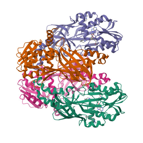
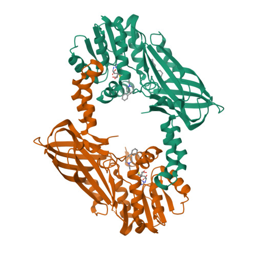
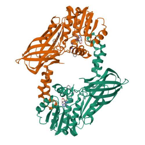
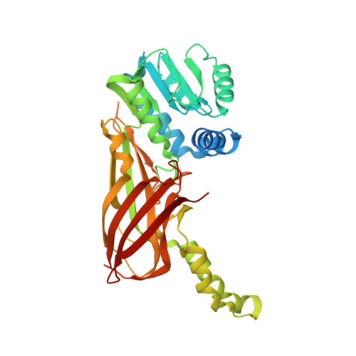
    5EGS

  • PubMed Abstract: 

    Protein methyltransferases (PMTs) are a promising target class in oncology and other disease areas. They are composed of SET domain methyltransferases and structurally unrelated Rossman-fold enzymes that include protein arginine methyltransferases (PRMTs). In the absence of a well-defined medicinal chemistry tool-kit focused on PMTs, most current inhibitors were identified by screening large and diverse libraries of leadlike molecules. So far, no successful fragment-based approach was reported against this target class. Here, by deconstructing potent PRMT inhibitors, we find that chemical moieties occupying the substrate arginine-binding site can act as efficient fragment inhibitors. Screening a fragment library against PRMT6 produced numerous hits, including a 300 nM inhibitor (ligand efficiency of 0.56) that decreased global histone 3 arginine 2 methylation in cells, and can serve as a warhead for the development of PRMT chemical probes.


  • Organizational Affiliation

    Structural Genomics Consortium, University of Toronto , Toronto, ON M5G 1L7, Canada.


Macromolecules
Find similar proteins by:  (by identity cutoff)  |  3D Structure
Entity ID: 1
MoleculeChains Sequence LengthOrganismDetailsImage
Protein arginine N-methyltransferase 6
A, B, C, D
376Homo sapiensMutation(s): 0 
Gene Names: PRMT6HRMT1L6
EC: 2.1.1 (PDB Primary Data), 2.1.1.125 (PDB Primary Data), 2.1.1.319 (UniProt)
UniProt & NIH Common Fund Data Resources
Find proteins for Q96LA8 (Homo sapiens)
Explore Q96LA8 
Go to UniProtKB:  Q96LA8
PHAROS:  Q96LA8
GTEx:  ENSG00000198890 
Entity Groups  
Sequence Clusters30% Identity50% Identity70% Identity90% Identity95% Identity100% Identity
UniProt GroupQ96LA8
Sequence Annotations
Expand
  • Reference Sequence
Small Molecules
Ligands 2 Unique
IDChains Name / Formula / InChI Key2D Diagram3D Interactions
SAH
Query on SAH

Download Ideal Coordinates CCD File 
E [auth A],
H [auth B],
J [auth C],
M [auth D]
S-ADENOSYL-L-HOMOCYSTEINE
C14 H20 N6 O5 S
ZJUKTBDSGOFHSH-WFMPWKQPSA-N
5NR
Query on 5NR

Download Ideal Coordinates CCD File 
F [auth A]
G [auth A]
I [auth B]
K [auth C]
L [auth C]
2-[4-(phenylmethyl)piperidin-1-yl]ethanamine
C14 H22 N2
PCNDXYHWLSHXMV-UHFFFAOYSA-N
Binding Affinity Annotations 
IDSourceBinding Affinity
5NR BindingDB:  5EGS Kd: 970 (nM) from 1 assay(s)
IC50: min: 230, max: 2.10e+4 (nM) from 2 assay(s)
SAH BindingDB:  5EGS IC50: 2177 (nM) from 1 assay(s)
Experimental Data & Validation

Experimental Data

  • Method: X-RAY DIFFRACTION
  • Resolution: 2.15 Å
  • R-Value Free:  0.246 (Depositor), 0.250 (DCC) 
  • R-Value Work:  0.201 (Depositor), 0.210 (DCC) 
  • R-Value Observed: 0.202 (Depositor) 
Space Group: P 1 21 1
Unit Cell:
Length ( Å )Angle ( ˚ )
a = 77.157α = 90
b = 135.616β = 98.91
c = 83.086γ = 90
Software Package:
Software NamePurpose
REFMACrefinement
XDSdata reduction
XSCALEdata scaling
PHASERphasing

Structure Validation

View Full Validation Report


Ligand Structure Quality Assessment 

Created with Raphaël 2.3.0Worse 01 BetterLigand structure goodness of fit to experimental dataBest fitted SAHClick on this verticalbar to view detailsBest fitted 5NRClick on this verticalbar to view details

Entry History 

Deposition Data

Revision History  (Full details and data files)

  • Version 1.0: 2016-02-03
    Type: Initial release
  • Version 1.1: 2016-06-22
    Changes: Database references
  • Version 1.2: 2024-01-10
    Changes: Data collection, Database references, Refinement description