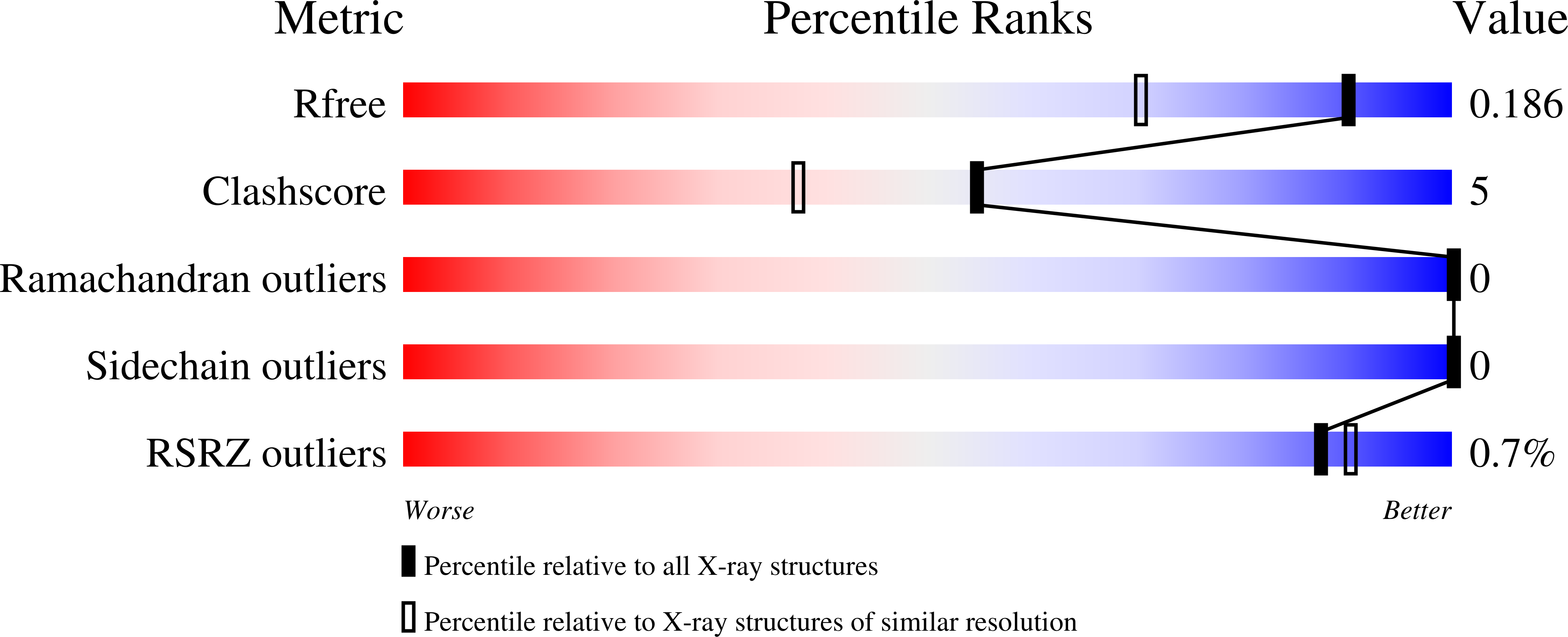
5E89

Crystal structure of Human galectin-3 CRD in complex with 3-fluophenyl-1,2,3-triazolyl thiodigalactoside inhibitor


Experimental Data Snapshot

  • Method: X-RAY DIFFRACTION
  • Resolution: 1.50 Å
  • R-Value Free: 0.190 
  • R-Value Work: 0.168 
  • R-Value Observed: 0.169 

Starting Model: experimental
View more details

wwPDB Validation   3D Report Full Report


Ligand Structure Quality Assessment 


This is version 1.2 of the entry. See complete history


Literature

Galectin-3-Binding Glycomimetics that Strongly Reduce Bleomycin-Induced Lung Fibrosis and Modulate Intracellular Glycan Recognition.

Delaine, T.Collins, P.MacKinnon, A.Sharma, G.Stegmayr, J.Rajput, V.K.Mandal, S.Cumpstey, I.Larumbe, A.Salameh, B.A.Kahl-Knutsson, B.van Hattum, H.van Scherpenzeel, M.Pieters, R.J.Sethi, T.Schambye, H.Oredsson, S.Leffler, H.Blanchard, H.Nilsson, U.J.

(2016) Chembiochem 17: 1759-1770

  • DOI: https://doi.org/10.1002/cbic.201600285
  • Primary Citation of Related Structures:  
    5E88, 5E89, 5E8A

  • PubMed Abstract: 

    Discovery of glycan-competitive galectin-3-binding compounds that attenuate lung fibrosis in a murine model and that block intracellular galectin-3 accumulation at damaged vesicles, hence revealing galectin-3-glycan interactions involved in fibrosis progression and in intracellular galectin-3 activities, is reported. 3,3'-Bis-(4-aryltriazol-1-yl)thiodigalactosides were synthesized and evaluated as antagonists of galectin-1, -2, -3, and -4 N-terminal, -4 C-terminal, -7 and -8 N-terminal, -9 N-terminal, and -9 C-terminal domains. Compounds displaying low-nanomolar affinities for galectins-1 and -3 were identified in a competitive fluorescence anisotropy assay. X-ray structural analysis of selected compounds in complex with galectin-3, together with galectin-3 mutant binding experiments, revealed that both the aryltriazolyl moieties and fluoro substituents on the compounds are involved in key interactions responsible for exceptional affinities towards galectin-3. The most potent galectin-3 antagonist was demonstrated to act in an assay monitoring galectin-3 accumulation upon amitriptyline-induced vesicle damage, visualizing a biochemically/medically relevant intracellular lectin-carbohydrate binding event and that it can be blocked by a small molecule. The same antagonist administered intratracheally attenuated bleomycin-induced pulmonary fibrosis in a mouse model with a dose/response profile comparing favorably with that of oral administration of the marketed antifibrotic compound pirfenidone.


  • Organizational Affiliation

    Centre for Analysis and Synthesis, Department of Chemistry, Lund University, P. O. Box 124, 221 00, Lund, Sweden.


Macromolecules
Find similar proteins by:  (by identity cutoff)  |  3D Structure
Entity ID: 1
MoleculeChains Sequence LengthOrganismDetailsImage
Galectin-3139Homo sapiensMutation(s): 0 
Gene Names: LGALS3MAC2
UniProt & NIH Common Fund Data Resources
Find proteins for P17931 (Homo sapiens)
Explore P17931 
Go to UniProtKB:  P17931
PHAROS:  P17931
GTEx:  ENSG00000131981 
Entity Groups  
Sequence Clusters30% Identity50% Identity70% Identity90% Identity95% Identity100% Identity
UniProt GroupP17931
Sequence Annotations
Expand
  • Reference Sequence
Small Molecules
Ligands 2 Unique
IDChains Name / Formula / InChI Key2D Diagram3D Interactions
TD2
Query on TD2

Download Ideal Coordinates CCD File 
C [auth A]3-deoxy-3-[4-(3-fluorophenyl)-1H-1,2,3-triazol-1-yl]-beta-D-galactopyranosyl 3-deoxy-3-[4-(3-fluorophenyl)-1H-1,2,3-triazol-1-yl]-1-thio-beta-D-galactopyranoside
C28 H30 F2 N6 O8 S
YGIDGBAHDZEYMT-MQFIMZJJSA-N
CL
Query on CL

Download Ideal Coordinates CCD File 
B [auth A]CHLORIDE ION
Cl
VEXZGXHMUGYJMC-UHFFFAOYSA-M
Binding Affinity Annotations 
IDSourceBinding Affinity
TD2 BindingDB:  5E89 Kd: min: 2.2, max: 37 (nM) from 4 assay(s)
IC50: min: 10, max: 300 (nM) from 4 assay(s)
Experimental Data & Validation

Experimental Data

  • Method: X-RAY DIFFRACTION
  • Resolution: 1.50 Å
  • R-Value Free: 0.190 
  • R-Value Work: 0.168 
  • R-Value Observed: 0.169 
  • Space Group: P 21 21 21
Unit Cell:
Length ( Å )Angle ( ˚ )
a = 37.045α = 90
b = 58.298β = 90
c = 63.728γ = 90
Software Package:
Software NamePurpose
SAINTdata reduction
SCALAdata scaling
REFMACrefinement
PDB_EXTRACTdata extraction
REFMACphasing

Structure Validation

View Full Validation Report


Ligand Structure Quality Assessment 


Entry History 

Deposition Data

Revision History  (Full details and data files)

  • Version 1.0: 2016-08-24
    Type: Initial release
  • Version 1.1: 2016-09-21
    Changes: Database references
  • Version 1.2: 2023-09-27
    Changes: Data collection, Database references, Derived calculations, Refinement description