5CEH

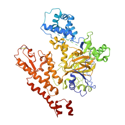
Structure of histone lysine demethylase KDM5A in complex with selective inhibitor


Experimental Data Snapshot

  • Method: X-RAY DIFFRACTION
  • Resolution: 3.14 Å
  • R-Value Free: 0.238 
  • R-Value Work: 0.221 
  • R-Value Observed: 0.222 

wwPDB Validation   3D Report Full Report


Ligand Structure Quality Assessment 


This is version 1.5 of the entry. See complete history


Literature

An inhibitor of KDM5 demethylases reduces survival of drug-tolerant cancer cells.

Vinogradova, M.Gehling, V.S.Gustafson, A.Arora, S.Tindell, C.A.Wilson, C.Williamson, K.E.Guler, G.D.Gangurde, P.Manieri, W.Busby, J.Flynn, E.M.Lan, F.Kim, H.J.Odate, S.Cochran, A.G.Liu, Y.Wongchenko, M.Yang, Y.Cheung, T.K.Maile, T.M.Lau, T.Costa, M.Hegde, G.V.Jackson, E.Pitti, R.Arnott, D.Bailey, C.Bellon, S.Cummings, R.T.Albrecht, B.K.Harmange, J.C.Kiefer, J.R.Trojer, P.Classon, M.

(2016) Nat Chem Biol 12: 531-538

  • DOI: https://doi.org/10.1038/nchembio.2085
  • Primary Citation of Related Structures:  
    5CEH

  • PubMed Abstract: 

    The KDM5 family of histone demethylases catalyzes the demethylation of histone H3 on lysine 4 (H3K4) and is required for the survival of drug-tolerant persister cancer cells (DTPs). Here we report the discovery and characterization of the specific KDM5 inhibitor CPI-455. The crystal structure of KDM5A revealed the mechanism of inhibition of CPI-455 as well as the topological arrangements of protein domains that influence substrate binding. CPI-455 mediated KDM5 inhibition, elevated global levels of H3K4 trimethylation (H3K4me3) and decreased the number of DTPs in multiple cancer cell line models treated with standard chemotherapy or targeted agents. These findings show that pretreatment of cancer cells with a KDM5-specific inhibitor results in the ablation of a subpopulation of cancer cells that can serve as the founders for therapeutic relapse.


  • Organizational Affiliation

    Genentech Inc., South San Francisco, California, USA.


Macromolecules
Find similar proteins by:  (by identity cutoff)  |  3D Structure
Entity ID: 1
MoleculeChains Sequence LengthOrganismDetailsImage
Lysine-specific demethylase 5A790Homo sapiensMutation(s): 0 
Gene Names: KDM5AJARID1ARBBP2RBP2
EC: 1.14.11 (PDB Primary Data), 1.14.11.67 (UniProt)
UniProt & NIH Common Fund Data Resources
Find proteins for P29375 (Homo sapiens)
Explore P29375 
Go to UniProtKB:  P29375
PHAROS:  P29375
GTEx:  ENSG00000073614 
Entity Groups  
Sequence Clusters30% Identity50% Identity70% Identity90% Identity95% Identity100% Identity
UniProt GroupP29375
Sequence Annotations
Expand
  • Reference Sequence

Find similar proteins by:  Sequence   |   3D Structure  

Entity ID: 2
MoleculeChains Sequence LengthOrganismDetailsImage
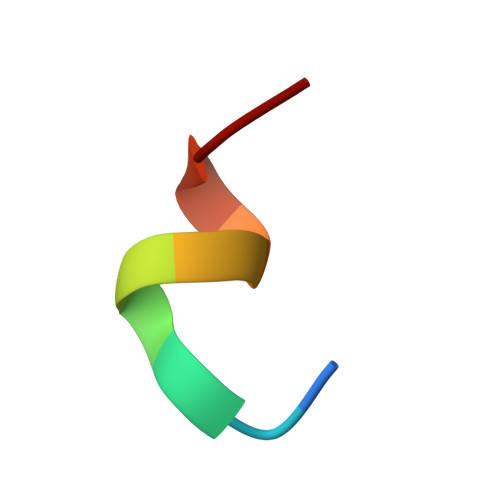
Unknown Peptide10Homo sapiensMutation(s): 0 
Gene Names: KDM5AJARID1ARBBP2RBP2
Sequence Annotations
Expand
  • Reference Sequence
Small Molecules
Binding Affinity Annotations 
IDSourceBinding Affinity
50P BindingDB:  5CEH IC50: min: 10, max: 506 (nM) from 3 assay(s)
EC50: 5200 (nM) from 1 assay(s)
Experimental Data & Validation

Experimental Data

  • Method: X-RAY DIFFRACTION
  • Resolution: 3.14 Å
  • R-Value Free: 0.238 
  • R-Value Work: 0.221 
  • R-Value Observed: 0.222 
  • Space Group: P 31 2 1
Unit Cell:
Length ( Å )Angle ( ˚ )
a = 159.306α = 90
b = 159.306β = 90
c = 92.462γ = 120
Software Package:
Software NamePurpose
PHENIXrefinement
HKL-2000data collection
SCALEPACKdata scaling
PHASERphasing
PDB_EXTRACTdata extraction

Structure Validation

View Full Validation Report


Ligand Structure Quality Assessment 


Entry History 

Deposition Data

Revision History  (Full details and data files)

  • Version 1.0: 2016-05-18
    Type: Initial release
  • Version 1.1: 2016-06-08
    Changes: Database references
  • Version 1.2: 2016-07-06
    Changes: Database references
  • Version 1.3: 2016-07-20
    Changes: Data collection
  • Version 1.4: 2017-11-22
    Changes: Derived calculations, Refinement description
  • Version 1.5: 2024-10-16
    Changes: Data collection, Database references, Derived calculations, Structure summary