5B5G

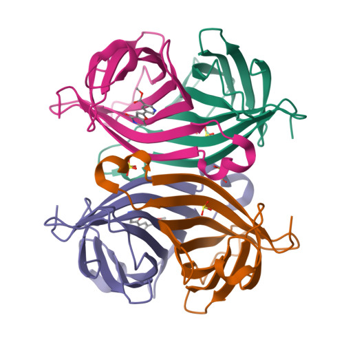
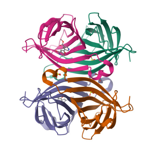
Crystal structure of ALiS4-Streptavidin complex


Experimental Data Snapshot

  • Method: X-RAY DIFFRACTION
  • Resolution: 1.50 Å
  • R-Value Free: 0.193 
  • R-Value Work: 0.145 
  • R-Value Observed: 0.147 

Starting Model: experimental
View more details

wwPDB Validation   3D Report Full Report


Ligand Structure Quality Assessment 


This is version 1.2 of the entry. See complete history


Literature

Improving the Solubility of Artificial Ligands of Streptavidin to Enable More Practical Reversible Switching of Protein Localization in Cells

Tachibana, R.Terai, T.Boncompain, G.Sugiyama, S.Saito, N.Perez, F.Urano, Y.

(2017) Chembiochem 18: 358-362

  • DOI: https://doi.org/10.1002/cbic.201600640
  • Primary Citation of Related Structures:  
    5B5F, 5B5G

  • PubMed Abstract: 

    Chemical inducers that can control target-protein localization in living cells are powerful tools to investigate dynamic biological systems. We recently reported the retention using selective hook or "RUSH" system for reversible localization change of proteins of interest by addition/washout of small-molecule artificial ligands of streptavidin (ALiS). However, the utility of previously developed ALiS was restricted by limited solubility in water. Here, we overcame this problem by X-ray crystal structure-guided design of a more soluble ALiS derivative (ALiS-3), which retains sufficient streptavidin-binding affinity for use in the RUSH system. The ALiS-3-streptavidin interaction was characterized in detail. ALiS-3 is a convenient and effective tool for dynamic control of α-mannosidase II localization between ER and Golgi in living cells.


  • Organizational Affiliation

    Graduate School of Pharmaceutical Sciences, The University of Tokyo, 7-3-1 Hongo, Bunkyo-ku, Tokyo, 113-0033, Japan.


Macromolecules
Find similar proteins by:  (by identity cutoff)  |  3D Structure
Entity ID: 1
MoleculeChains Sequence LengthOrganismDetailsImage
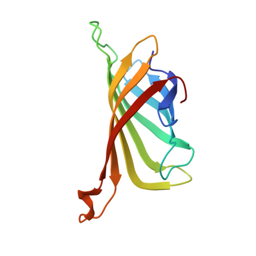
Streptavidin
A, B, C, D
120Streptomyces avidiniiMutation(s): 0 
UniProt
Find proteins for P22629 (Streptomyces avidinii)
Explore P22629 
Go to UniProtKB:  P22629
Entity Groups  
Sequence Clusters30% Identity50% Identity70% Identity90% Identity95% Identity100% Identity
UniProt GroupP22629
Sequence Annotations
Expand
  • Reference Sequence
Small Molecules
Ligands 2 Unique
IDChains Name / Formula / InChI Key2D Diagram3D Interactions
6FX
Query on 6FX

Download Ideal Coordinates CCD File 
K [auth C],
M [auth D]
methyl 5-(4-oxidanylidene-5~{H}-furo[3,2-c]pyridin-2-yl)pyridine-3-carboxylate
C14 H10 N2 O4
CKTWBGKZILYBCF-UHFFFAOYSA-N
SO3
Query on SO3

Download Ideal Coordinates CCD File 
E [auth A]
F [auth A]
G [auth A]
H [auth B]
I [auth B]
E [auth A],
F [auth A],
G [auth A],
H [auth B],
I [auth B],
J [auth C],
L [auth D]
SULFITE ION
O3 S
LSNNMFCWUKXFEE-UHFFFAOYSA-L
Experimental Data & Validation

Experimental Data

  • Method: X-RAY DIFFRACTION
  • Resolution: 1.50 Å
  • R-Value Free: 0.193 
  • R-Value Work: 0.145 
  • R-Value Observed: 0.147 
  • Space Group: P 1 21 1
Unit Cell:
Length ( Å )Angle ( ˚ )
a = 57.921α = 90
b = 84.423β = 98.98
c = 46.138γ = 90
Software Package:
Software NamePurpose
REFMACrefinement
HKL-2000data reduction
HKL-2000data scaling
MOLREPphasing

Structure Validation

View Full Validation Report


Ligand Structure Quality Assessment 


Entry History & Funding Information

Deposition Data


Funding OrganizationLocationGrant Number
JSPS KAKENHIJapan25286051
JSPS KAKENHIJapan25650051
JSPS KAKENHIJapan23860028

Revision History  (Full details and data files)

  • Version 1.0: 2017-03-01
    Type: Initial release
  • Version 1.1: 2020-02-26
    Changes: Data collection
  • Version 1.2: 2023-11-08
    Changes: Data collection, Database references, Refinement description