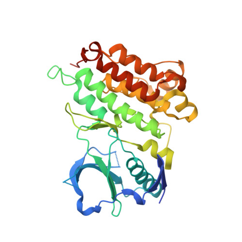
5AAB

Structure of C1156Y,L1198F Mutant Human Anaplastic Lymphoma Kinase in Complex with Crizotinib


Experimental Data Snapshot

  • Method: X-RAY DIFFRACTION
  • Resolution: 2.20 Å
  • R-Value Free: 0.246 
  • R-Value Work: 0.210 
  • R-Value Observed: 0.211 

Starting Model: experimental
View more details

wwPDB Validation   3D Report Full Report


Ligand Structure Quality Assessment 


This is version 1.2 of the entry. See complete history


Literature

Resensitization to Crizotinib by the Lorlatinib Alk Resistance Mutation L1198F.

Shaw, A.T.Friboulet, L.Leshchiner, I.Gainor, J.F.Bergqvist, S.Brooun, A.Burke, B.J.Deng, Y.Liu, W.Dardaei, L.Frias, R.L.Schultz, K.R.Logan, J.James, L.P.Smeal, T.Timofeevski, S.Katayama, R.Iafrate, A.J.Le, L.Mctigue, M.Getz, G.Johnson, T.W.Engelman, J.A.

(2016) N Engl J Med 374: 54

  • DOI: https://doi.org/10.1056/NEJMoa1508887
  • Primary Citation of Related Structures:  
    5A9U, 5AA8, 5AA9, 5AAA, 5AAB, 5AAC

  • PubMed Abstract: 

    In a patient who had metastatic anaplastic lymphoma kinase (ALK)-rearranged lung cancer, resistance to crizotinib developed because of a mutation in the ALK kinase domain. This mutation is predicted to result in a substitution of cysteine by tyrosine at amino acid residue 1156 (C1156Y). Her tumor did not respond to a second-generation ALK inhibitor, but it did respond to lorlatinib (PF-06463922), a third-generation inhibitor. When her tumor relapsed, sequencing of the resistant tumor revealed an ALK L1198F mutation in addition to the C1156Y mutation. The L1198F substitution confers resistance to lorlatinib through steric interference with drug binding. However, L1198F paradoxically enhances binding to crizotinib, negating the effect of C1156Y and resensitizing resistant cancers to crizotinib. The patient received crizotinib again, and her cancer-related symptoms and liver failure resolved. (Funded by Pfizer and others; ClinicalTrials.gov number, NCT01970865.).


  • Organizational Affiliation

    Massachusetts General Hospital Cancer Center (A.T.S., L.F., J.F.G., L.D., R.L.F., K.R.S., J.L., G.G., J.A.E.) and the Department of Pathology (A.J.I., L.L., G.G.), Massachusetts General Hospital, Boston, and the Broad Institute of the Massachusetts Institute of Technology and Harvard, Cambridge (I.L., G.G.) - all in Massachusetts; Pfizer Worldwide Research and Development, La Jolla, CA (S.B., A.B., B.J.B., Y.-L.D., W.L., L.P.J., T.S., S.T., M.M., T.W.J.); and the Japanese Foundation for Cancer Research, Tokyo (R.K.).


Macromolecules
Find similar proteins by:  (by identity cutoff)  |  3D Structure
Entity ID: 1
MoleculeChains Sequence LengthOrganismDetailsImage
ALK TYROSINE KINASE RECEPTOR327Homo sapiensMutation(s): 2 
EC: 2.7.10.1
UniProt & NIH Common Fund Data Resources
Find proteins for Q9UM73 (Homo sapiens)
Explore Q9UM73 
Go to UniProtKB:  Q9UM73
PHAROS:  Q9UM73
GTEx:  ENSG00000171094 
Entity Groups  
Sequence Clusters30% Identity50% Identity70% Identity90% Identity95% Identity100% Identity
UniProt GroupQ9UM73
Sequence Annotations
Expand
  • Reference Sequence
Small Molecules
Ligands 1 Unique
IDChains Name / Formula / InChI Key2D Diagram3D Interactions
VGH
Query on VGH

Download Ideal Coordinates CCD File 
B [auth A]3-[(1R)-1-(2,6-dichloro-3-fluorophenyl)ethoxy]-5-(1-piperidin-4-yl-1H-pyrazol-4-yl)pyridin-2-amine
C21 H22 Cl2 F N5 O
KTEIFNKAUNYNJU-GFCCVEGCSA-N
Binding Affinity Annotations 
IDSourceBinding Affinity
VGH BindingDB:  5AAB Ki: min: 0.74, max: 8.2 (nM) from 2 assay(s)
Kd: min: 3.3, max: 4.4 (nM) from 2 assay(s)
IC50: min: 0.64, max: 3039 (nM) from 79 assay(s)
EC50: min: 575, max: 2.28e+4 (nM) from 3 assay(s)
Experimental Data & Validation

Experimental Data

  • Method: X-RAY DIFFRACTION
  • Resolution: 2.20 Å
  • R-Value Free: 0.246 
  • R-Value Work: 0.210 
  • R-Value Observed: 0.211 
  • Space Group: P 21 21 21
Unit Cell:
Length ( Å )Angle ( ˚ )
a = 51.541α = 90
b = 57.449β = 90
c = 104.787γ = 90
Software Package:
Software NamePurpose
CNXrefinement
autoPROCdata reduction
Aimlessdata scaling
CNXphasing

Structure Validation

View Full Validation Report


Ligand Structure Quality Assessment 


Entry History 

Deposition Data

Revision History  (Full details and data files)

  • Version 1.0: 2016-06-08
    Type: Initial release
  • Version 1.1: 2019-05-08
    Changes: Data collection, Experimental preparation, Other
  • Version 1.2: 2024-01-10
    Changes: Data collection, Database references, Derived calculations, Other, Refinement description