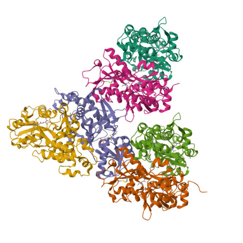
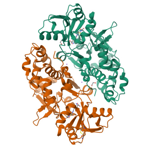
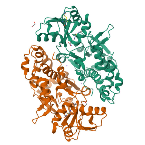
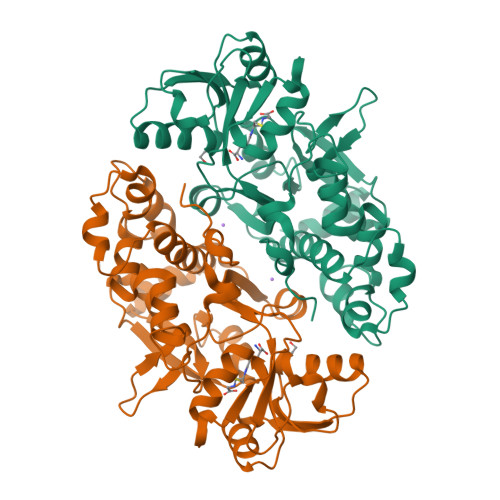
ABC transporter / periplasmic binding protein from Brucella ovis with glutathione bound
Seattle Structural Genomics Center for Infectious Disease (SSGCID)To be published.
Experimental Data Snapshot
Entity ID: 1 | |||||
|---|---|---|---|---|---|
| Molecule | Chains | Sequence Length | Organism | Details | Image |
| Amino acid ABC transporter, periplasmic amino acid-binding protein | 328 | Brucella ovis ATCC 25840 | Mutation(s): 0 Gene Names: BOV_0736 | 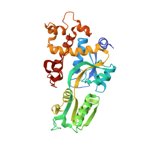 | |
UniProt | |||||
Find proteins for A0A0M3KL33 (Brucella ovis (strain ATCC 25840 / 63/290 / NCTC 10512)) Explore A0A0M3KL33 Go to UniProtKB: A0A0M3KL33 | |||||
Entity Groups | |||||
| Sequence Clusters | 30% Identity50% Identity70% Identity90% Identity95% Identity100% Identity | ||||
| UniProt Group | A0A0M3KL33 | ||||
Sequence AnnotationsExpand | |||||
| |||||
| Ligands 3 Unique | |||||
|---|---|---|---|---|---|
| ID | Chains | Name / Formula / InChI Key | 2D Diagram | 3D Interactions | |
| GSH Query on GSH | G [auth A] L [auth B] Q [auth C] U [auth D] W [auth E] | GLUTATHIONE C10 H17 N3 O6 S RWSXRVCMGQZWBV-WDSKDSINSA-N |  | ||
| EDO Query on EDO | I [auth A] J [auth A] N [auth B] O [auth B] S [auth C] | 1,2-ETHANEDIOL C2 H6 O2 LYCAIKOWRPUZTN-UHFFFAOYSA-N |  | ||
| NA Query on NA | H [auth A] K [auth A] M [auth B] P [auth B] R [auth C] | SODIUM ION Na FKNQFGJONOIPTF-UHFFFAOYSA-N |  | ||
| Length ( Å ) | Angle ( ˚ ) |
|---|---|
| a = 93.99 | α = 90 |
| b = 76.93 | β = 94.07 |
| c = 131.47 | γ = 90 |
| Software Name | Purpose |
|---|---|
| PHENIX | refinement |
| XDS | data reduction |
| XSCALE | data scaling |
| PHENIX | phasing |
RCSB PDB is hosted by

RCSB PDB is a member of the