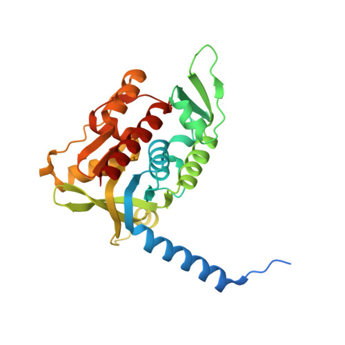
4Y4N

Thiazole synthase Thi4 from Methanococcus igneus


Experimental Data Snapshot

  • Method: X-RAY DIFFRACTION
  • Resolution: 2.10 Å
  • R-Value Free: 0.216 
  • R-Value Work: 0.199 
  • R-Value Observed: 0.200 

Starting Model: experimental
View more details

wwPDB Validation   3D Report Full Report


Ligand Structure Quality Assessment 


This is version 1.3 of the entry. See complete history


Literature

Structural Basis for Iron-Mediated Sulfur Transfer in Archael and Yeast Thiazole Synthases.

Zhang, X.Eser, B.E.Chanani, P.K.Begley, T.P.Ealick, S.E.

(2016) Biochemistry 55: 1826-1838

  • DOI: https://doi.org/10.1021/acs.biochem.6b00030
  • Primary Citation of Related Structures:  
    4Y4L, 4Y4M, 4Y4N

  • PubMed Abstract: 

    Thiamin diphosphate is an essential cofactor in all forms of life and plays a key role in amino acid and carbohydrate metabolism. Its biosynthesis involves separate syntheses of the pyrimidine and thiazole moieties, which are then coupled to form thiamin monophosphate. A final phosphorylation produces the active form of the cofactor. In most bacteria, six gene products are required for biosynthesis of the thiamin thiazole. In yeast and fungi only one gene product, Thi4, is required for thiazole biosynthesis. Methanococcus jannaschii expresses a putative Thi4 ortholog that was previously reported to be a ribulose 1,5-bisphosphate synthase [Finn, M. W. and Tabita, F. R. (2004) J. Bacteriol., 186, 6360-6366]. Our structural studies show that the Thi4 orthologs from M. jannaschii and Methanococcus igneus are structurally similar to Thi4 from Saccharomyces cerevisiae. In addition, all active site residues are conserved except for a key cysteine residue, which in S. cerevisiae is the source of the thiazole sulfur atom. Our recent biochemical studies showed that the archael Thi4 orthologs use nicotinamide adenine dinucleotide, glycine, and free sulfide to form the thiamin thiazole in an iron-dependent reaction [Eser, B., Zhang, X., Chanani, P. K., Begley, T. P., and Ealick, S. E. (2016) J. Am. Chem. Soc. , DOI: 10.1021/jacs.6b00445]. Here we report X-ray crystal structures of Thi4 from M. jannaschii complexed with ADP-ribulose, the C205S variant of Thi4 from S. cerevisiae with a bound glycine imine intermediate, and Thi4 from M. igneus with bound glycine imine intermediate and iron. These studies reveal the structural basis for the iron-dependent mechanism of sulfur transfer in archael and yeast thiazole synthases.


  • Organizational Affiliation

    Department of Chemistry and Chemical Biology, Cornell University , Ithaca, New York 14853, United States.


Macromolecules
Find similar proteins by:  (by identity cutoff)  |  3D Structure
Entity ID: 1
MoleculeChains Sequence LengthOrganismDetailsImage
Putative ribose 1,5-bisphosphate isomerase
A, B, C, D, E
A, B, C, D, E, F, G, H
286Methanotorris igneus Kol 5Mutation(s): 0 
Gene Names: Metig_0735
EC: 5.3.1.29 (PDB Primary Data), 2.4.2.59 (UniProt)
UniProt
Find proteins for F6BCS4 (Methanotorris igneus (strain DSM 5666 / JCM 11834 / Kol 5))
Explore F6BCS4 
Go to UniProtKB:  F6BCS4
Entity Groups  
Sequence Clusters30% Identity50% Identity70% Identity90% Identity95% Identity100% Identity
UniProt GroupF6BCS4
Sequence Annotations
Expand
  • Reference Sequence
Small Molecules
Ligands 2 Unique
IDChains Name / Formula / InChI Key2D Diagram3D Interactions
48H
Query on 48H

Download Ideal Coordinates CCD File 
J [auth A]
L [auth A]
N [auth B]
P [auth B]
R [auth C]
J [auth A],
L [auth A],
N [auth B],
P [auth B],
R [auth C],
T [auth D],
V [auth E],
X [auth F]
2-[(E)-[(4R)-5-[[[(2R,3S,4R,5R)-5-(6-aminopurin-9-yl)-3,4-bis(oxidanyl)oxolan-2-yl]methoxy-oxidanyl-phosphoryl]oxy-oxidanyl-phosphoryl]oxy-4-oxidanyl-3-oxidanylidene-pentan-2-ylidene]amino]ethanoic acid
C17 H24 N6 O14 P2
IOANXAZHBCZCOH-NTPWJJDISA-N
FE2
Query on FE2

Download Ideal Coordinates CCD File 
I [auth A]
K [auth A]
M [auth B]
O [auth B]
Q [auth C]
I [auth A],
K [auth A],
M [auth B],
O [auth B],
Q [auth C],
S [auth D],
U [auth E],
W [auth F]
FE (II) ION
Fe
CWYNVVGOOAEACU-UHFFFAOYSA-N
Experimental Data & Validation

Experimental Data

  • Method: X-RAY DIFFRACTION
  • Resolution: 2.10 Å
  • R-Value Free: 0.216 
  • R-Value Work: 0.199 
  • R-Value Observed: 0.200 
  • Space Group: P 31
Unit Cell:
Length ( Å )Angle ( ˚ )
a = 101.302α = 90
b = 101.302β = 90
c = 204.309γ = 120
Software Package:
Software NamePurpose
REFMACrefinement
HKL-2000data reduction
HKL-2000data scaling
PHENIXphasing

Structure Validation

View Full Validation Report


Ligand Structure Quality Assessment 


Entry History 

Deposition Data

Revision History  (Full details and data files)

  • Version 1.0: 2016-03-09
    Type: Initial release
  • Version 1.1: 2016-05-04
    Changes: Database references
  • Version 1.2: 2023-09-27
    Changes: Data collection, Database references, Derived calculations, Refinement description
  • Version 1.3: 2024-11-06
    Changes: Structure summary