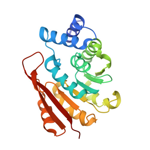
4XUE

Synthesis and evaluation of heterocyclic catechol mimics as inhibitors of catechol-O-methyltransferase (COMT): Structure with Cmpd27b


Experimental Data Snapshot

  • Method: X-RAY DIFFRACTION
  • Resolution: 2.30 Å
  • R-Value Free: 0.229 
  • R-Value Work: 0.198 
  • R-Value Observed: 0.200 

wwPDB Validation   3D Report Full Report


Ligand Structure Quality Assessment 


This is version 1.2 of the entry. See complete history


Literature

Synthesis and Evaluation of Heterocyclic Catechol Mimics as Inhibitors of Catechol-O-methyltransferase (COMT).

Harrison, S.T.Poslusney, M.S.Mulhearn, J.J.Zhao, Z.Kett, N.R.Schubert, J.W.Melamed, J.Y.Allison, T.J.Patel, S.B.Sanders, J.M.Sharma, S.Smith, R.F.Hall, D.L.Robinson, R.G.Sachs, N.A.Hutson, P.H.Wolkenberg, S.E.Barrow, J.C.

(2015) ACS Med Chem Lett 6: 318-323

  • DOI: https://doi.org/10.1021/ml500502d
  • Primary Citation of Related Structures:  
    4XUC, 4XUD, 4XUE

  • PubMed Abstract: 

    3-Hydroxy-4-pyridinones and 5-hydroxy-4-pyrimidinones were identified as inhibitors of catechol-O-methyltransferase (COMT) in a high-throughput screen. These heterocyclic catechol mimics exhibit potent inhibition of the enzyme and an improved toxicity profile versus the marketed nitrocatechol inhibitors tolcapone and entacapone. Optimization of the series was aided by X-ray cocrystal structures of the novel inhibitors in complex with COMT and cofactors SAM and Mg(2+). The crystal structures suggest a mechanism of inhibition for these heterocyclic inhibitors distinct from previously disclosed COMT inhibitors.


  • Organizational Affiliation

    Department of Medicinal Chemistry, Global Structural Biology, Chemical Modeling and Informatics, and Department of Neuroscience Research, Merck Research Laboratories , Sumneytown Pike, West Point, Pennsylvania 19486, United States.


Macromolecules
Find similar proteins by:  (by identity cutoff)  |  3D Structure
Entity ID: 1
MoleculeChains Sequence LengthOrganismDetailsImage
Catechol O-methyltransferaseA [auth B],
B [auth A]
215Homo sapiensMutation(s): 0 
Gene Names: COMT
EC: 2.1.1.6
UniProt & NIH Common Fund Data Resources
Find proteins for P21964 (Homo sapiens)
Explore P21964 
Go to UniProtKB:  P21964
PHAROS:  P21964
GTEx:  ENSG00000093010 
Entity Groups  
Sequence Clusters30% Identity50% Identity70% Identity90% Identity95% Identity100% Identity
UniProt GroupP21964
Sequence Annotations
Expand
  • Reference Sequence
Small Molecules
Ligands 3 Unique
IDChains Name / Formula / InChI Key2D Diagram3D Interactions
SAM
Query on SAM

Download Ideal Coordinates CCD File 
E [auth B],
H [auth A]
S-ADENOSYLMETHIONINE
C15 H22 N6 O5 S
MEFKEPWMEQBLKI-FCKMPRQPSA-N
43J
Query on 43J

Download Ideal Coordinates CCD File 
C [auth B],
F [auth A]
2-(biphenyl-3-yl)-5-hydroxy-3-methylpyrimidin-4(3H)-one
C17 H14 N2 O2
XFQLJHLLJPLAPL-UHFFFAOYSA-N
MG
Query on MG

Download Ideal Coordinates CCD File 
D [auth B],
G [auth A]
MAGNESIUM ION
Mg
JLVVSXFLKOJNIY-UHFFFAOYSA-N
Binding Affinity Annotations 
IDSourceBinding Affinity
43J BindingDB:  4XUE IC50: min: 170, max: 5000 (nM) from 2 assay(s)
Experimental Data & Validation

Experimental Data

  • Method: X-RAY DIFFRACTION
  • Resolution: 2.30 Å
  • R-Value Free: 0.229 
  • R-Value Work: 0.198 
  • R-Value Observed: 0.200 
  • Space Group: P 21 21 21
Unit Cell:
Length ( Å )Angle ( ˚ )
a = 55.043α = 90
b = 113.19β = 90
c = 113.818γ = 90
Software Package:
Software NamePurpose
SCALEPACKdata scaling
REFMACrefinement
PDB_EXTRACTdata extraction
MOLREPphasing

Structure Validation

View Full Validation Report


Ligand Structure Quality Assessment 


Entry History 

Deposition Data

Revision History  (Full details and data files)

  • Version 1.0: 2015-04-15
    Type: Initial release
  • Version 1.1: 2017-11-22
    Changes: Derived calculations, Refinement description, Source and taxonomy, Structure summary
  • Version 1.2: 2024-02-28
    Changes: Data collection, Database references, Derived calculations