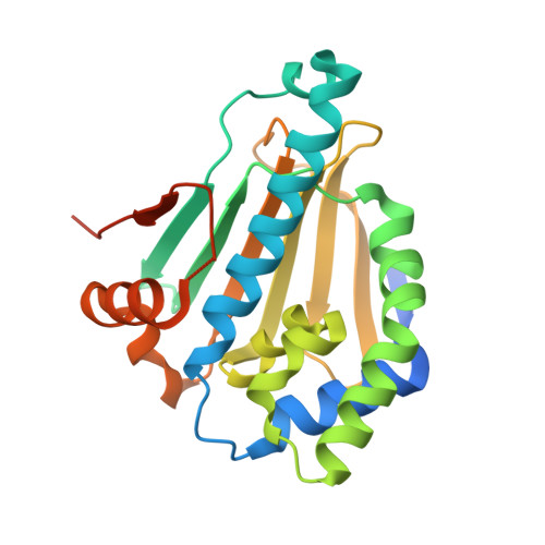
4NH7

Correlation between chemotype-dependent binding conformations of HSP90 alpha/beta and isoform selectivity


Experimental Data Snapshot

  • Method: X-RAY DIFFRACTION
  • Resolution: 2.00 Å
  • R-Value Free: 0.243 
  • R-Value Work: 0.206 
  • R-Value Observed: 0.208 

wwPDB Validation   3D Report Full Report


Ligand Structure Quality Assessment 


This is version 2.1 of the entry. See complete history


Literature

Correlation between chemotype-dependent binding conformations of HSP90 alpha / beta and isoform selectivity-Implications for the structure-based design of HSP90 alpha / beta selective inhibitors for treating neurodegenerative diseases.

Ernst, J.T.Liu, M.Zuccola, H.Neubert, T.Beaumont, K.Turnbull, A.Kallel, A.Vought, B.Stamos, D.

(2014) Bioorg Med Chem Lett 24: 204-208

  • DOI: https://doi.org/10.1016/j.bmcl.2013.11.036
  • Primary Citation of Related Structures:  
    4NH7, 4NH8, 4NH9

  • PubMed Abstract: 

    HSP90 continues to be a target of interest for neurodegeneration indications. Selective knockdown of the HSP90 cytosolic isoforms α and β is sufficient to reduce mutant huntingtin protein levels in vitro. Chemotype-dependent binding conformations of HSP90α/β appear to strongly influence isoform selectivity. The rational design of HSP90α/β inhibitors selective versus the mitochondrial (TRAP1) and endoplasmic reticulum (GRP94) isoforms offers a potential mitigating strategy for mechanism-based toxicities. Better tolerated HSP90 inhibitors would be attractive for targeting chronic neurodegenerative diseases such as Huntington's disease.


  • Organizational Affiliation

    Vertex Pharmaceuticals, Department of Chemistry and Drug Innovation, 11010 Torreyana Road, San Diego, CA 92121, United States. Electronic address: [email protected].


Macromolecules
Find similar proteins by:  (by identity cutoff)  |  3D Structure
Entity ID: 1
MoleculeChains Sequence LengthOrganismDetailsImage
Heat shock protein HSP 90-alpha
A, B
233Homo sapiensMutation(s): 0 
Gene Names: HSP90AA1HSP90AHSPC1HSPCA
EC: 3.6.4.10
UniProt & NIH Common Fund Data Resources
Find proteins for P07900 (Homo sapiens)
Explore P07900 
Go to UniProtKB:  P07900
PHAROS:  P07900
GTEx:  ENSG00000080824 
Entity Groups  
Sequence Clusters30% Identity50% Identity70% Identity90% Identity95% Identity100% Identity
UniProt GroupP07900
Sequence Annotations
Expand
  • Reference Sequence
Small Molecules
Ligands 2 Unique
IDChains Name / Formula / InChI Key2D Diagram3D Interactions
E0G
Query on E0G

Download Ideal Coordinates CCD File 
C [auth A],
G [auth B]
4-[6,6-dimethyl-4-oxidanylidene-3-(trifluoromethyl)-5,7-dihydroindazol-1-yl]-2-[(4-oxidanylcyclohexyl)amino]benzamide
C23 H27 F3 N4 O3
ZFVRYNYOPQZKDG-MQMHXKEQSA-N
GOL
Query on GOL

Download Ideal Coordinates CCD File 
D [auth A],
E [auth A],
F [auth A],
H [auth B],
I [auth B]
GLYCEROL
C3 H8 O3
PEDCQBHIVMGVHV-UHFFFAOYSA-N
Binding Affinity Annotations 
IDSourceBinding Affinity
E0G BindingDB:  4NH7 Kd: 4 (nM) from 1 assay(s)
IC50: min: 1, max: 1.00e+4 (nM) from 7 assay(s)
Experimental Data & Validation

Experimental Data

  • Method: X-RAY DIFFRACTION
  • Resolution: 2.00 Å
  • R-Value Free: 0.243 
  • R-Value Work: 0.206 
  • R-Value Observed: 0.208 
  • Space Group: C 1 2 1
Unit Cell:
Length ( Å )Angle ( ˚ )
a = 114.98α = 90
b = 88.04β = 124.53
c = 71.07γ = 90
Software Package:
Software NamePurpose
PROCESSdata collection
BUSTERrefinement
PROCESSdata reduction
PROCESSdata scaling
BUSTERphasing

Structure Validation

View Full Validation Report


Ligand Structure Quality Assessment 


Entry History 

Deposition Data

Revision History  (Full details and data files)

  • Version 1.0: 2014-01-15
    Type: Initial release
  • Version 2.0: 2017-11-29
    Changes: Atomic model, Database references, Derived calculations, Non-polymer description, Source and taxonomy, Structure summary
  • Version 2.1: 2024-02-28
    Changes: Data collection, Database references, Derived calculations