RCSB PDB - 4MTO: Structural Basis for Ca2+ Selectivity of a Voltage-gated Calcium Channel

 4MTO

Structural Basis for Ca2+ Selectivity of a Voltage-gated Calcium Channel


Experimental Data Snapshot

  • Method: X-RAY DIFFRACTION
  • Resolution: 3.40 Å
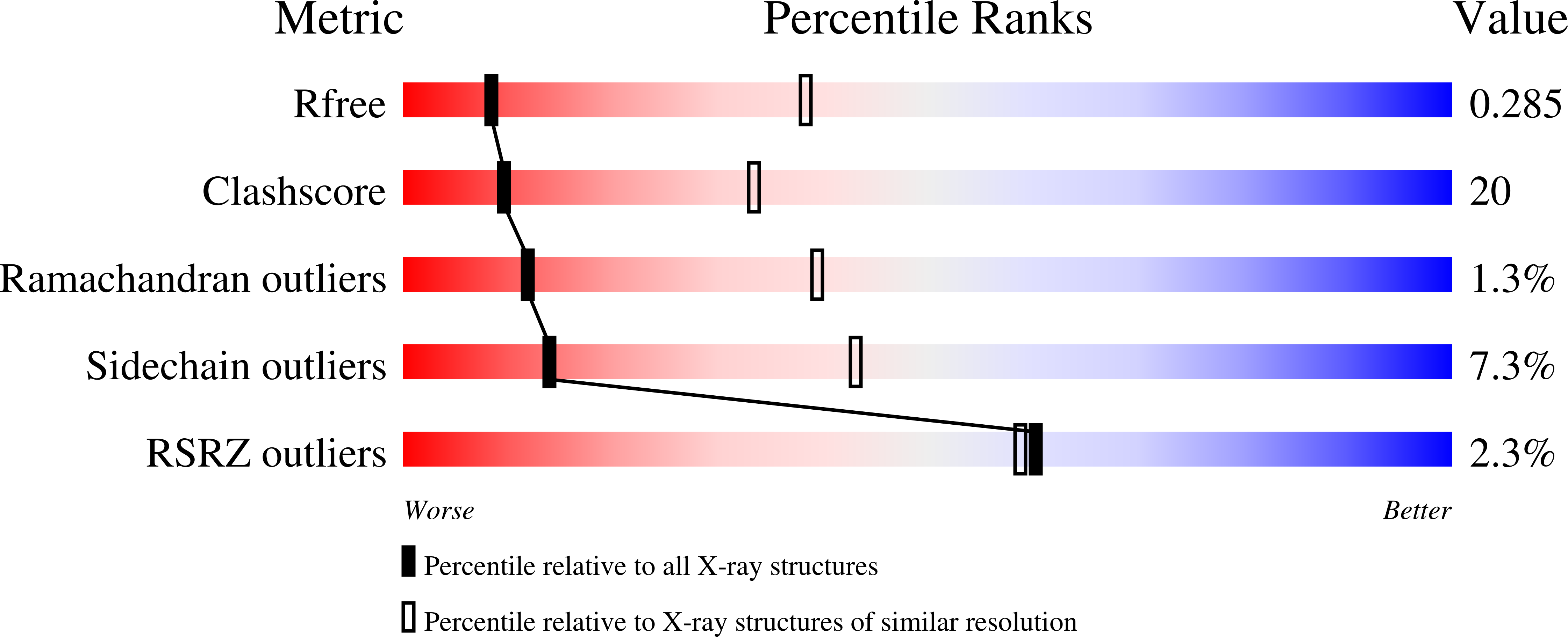
  • R-Value Free: 0.297 
  • R-Value Work: 0.260 
  • R-Value Observed: 0.261 

wwPDB Validation   3D Report Full Report


Ligand Structure Quality Assessment 

Created with Raphaël 2.3.0Worse 01 BetterLigand structure goodness of fit to experimental dataBest fitted PX4Click on this verticalbar to view details

This is version 1.4 of the entry. See complete history


Literature

Structural basis for Ca2+ selectivity of a voltage-gated calcium channel.

Tang, L.Gamal El-Din, T.M.Payandeh, J.Martinez, G.Q.Heard, T.M.Scheuer, T.Zheng, N.Catterall, W.A.

(2014) Nature 505: 56-61

  • DOI: https://doi.org/10.1038/nature12775
  • Primary Citation of Related Structures:  
    4MS2, 4MTF, 4MTG, 4MTO, 4MVM, 4MVO, 4MVQ, 4MVR, 4MVS, 4MVU, 4MVZ, 4MW3, 4MW8

  • PubMed Abstract: 

    Voltage-gated calcium (CaV) channels catalyse rapid, highly selective influx of Ca(2+) into cells despite a 70-fold higher extracellular concentration of Na(+). How CaV channels solve this fundamental biophysical problem remains unclear. Here we report physiological and crystallographic analyses of a calcium selectivity filter constructed in the homotetrameric bacterial NaV channel NaVAb. Our results reveal interactions of hydrated Ca(2+) with two high-affinity Ca(2+)-binding sites followed by a third lower-affinity site that would coordinate Ca(2+) as it moves inward. At the selectivity filter entry, Site 1 is formed by four carboxyl side chains, which have a critical role in determining Ca(2+) selectivity. Four carboxyls plus four backbone carbonyls form Site 2, which is targeted by the blocking cations Cd(2+) and Mn(2+), with single occupancy. The lower-affinity Site 3 is formed by four backbone carbonyls alone, which mediate exit into the central cavity. This pore architecture suggests a conduction pathway involving transitions between two main states with one or two hydrated Ca(2+) ions bound in the selectivity filter and supports a 'knock-off' mechanism of ion permeation through a stepwise-binding process. The multi-ion selectivity filter of our CaVAb model establishes a structural framework for understanding the mechanisms of ion selectivity and conductance by vertebrate CaV channels.


  • Organizational Affiliation

    1] Department of Pharmacology, University of Washington, Seattle, Washington 98195, USA [2] Howard Hughes Medical Institute, University of Washington, Seattle, Washington 98195, USA [3].


Macromolecules
Find similar proteins by:  (by identity cutoff)  |  3D Structure
Entity ID: 1
MoleculeChains Sequence LengthOrganismDetailsImage
Ion transport protein
A, B, C, D
237Aliarcobacter butzleri RM4018Mutation(s): 0 
Gene Names: Abu_1752
Membrane Entity: Yes 
UniProt
Find proteins for A8EVM5 (Aliarcobacter butzleri (strain RM4018))
Explore A8EVM5 
Go to UniProtKB:  A8EVM5
Entity Groups  
Sequence Clusters30% Identity50% Identity70% Identity90% Identity95% Identity100% Identity
UniProt GroupA8EVM5
Sequence Annotations
Expand
  • Reference Sequence
Small Molecules
Ligands 2 Unique
IDChains Name / Formula / InChI Key2D Diagram3D Interactions
PX4
Query on PX4

Download Ideal Coordinates CCD File 
G [auth A]
H [auth A]
I [auth A]
J [auth A]
K [auth A]
1,2-DIMYRISTOYL-SN-GLYCERO-3-PHOSPHOCHOLINE
C36 H73 N O8 P
CITHEXJVPOWHKC-UUWRZZSWSA-O
CA
Query on CA

Download Ideal Coordinates CCD File 
E [auth A],
F [auth A],
L [auth B]
CALCIUM ION
Ca
BHPQYMZQTOCNFJ-UHFFFAOYSA-N
Experimental Data & Validation

Experimental Data

  • Method: X-RAY DIFFRACTION
  • Resolution: 3.40 Å
  • R-Value Free: 0.297 
  • R-Value Work: 0.260 
  • R-Value Observed: 0.261 
  • Space Group: C 1 2 1
Unit Cell:
Length ( Å )Angle ( ˚ )
a = 176.635α = 90
b = 177.655β = 132.53
c = 130.6γ = 90
Software Package:
Software NamePurpose
HKL-2000data collection
PHENIXmodel building
REFMACrefinement
MOSFLMdata reduction
SCALAdata scaling
PHENIXphasing

Structure Validation

View Full Validation Report


Ligand Structure Quality Assessment 

Created with Raphaël 2.3.0Worse 01 BetterLigand structure goodness of fit to experimental dataBest fitted PX4Click on this verticalbar to view details

Entry History 

Deposition Data

Revision History  (Full details and data files)

  • Version 1.0: 2013-11-27
    Type: Initial release
  • Version 1.1: 2013-12-18
    Changes: Database references
  • Version 1.2: 2014-01-15
    Changes: Database references
  • Version 1.3: 2014-11-12
    Changes: Structure summary
  • Version 1.4: 2024-02-28
    Changes: Data collection, Database references, Derived calculations