4EP7

Functional implications from the Cid1 poly(U) polymerase crystal structure


Experimental Data Snapshot

  • Method: X-RAY DIFFRACTION
  • Resolution: 2.28 Å
  • R-Value Free: 
    0.247 (Depositor), 0.240 (DCC) 
  • R-Value Work: 
    0.188 (Depositor), 0.180 (DCC) 
  • R-Value Observed: 
    0.191 (Depositor) 

wwPDB Validation   3D Report Full Report


Ligand Structure Quality Assessment 

Created with Raphaël 2.3.0Worse 01 BetterLigand structure goodness of fit to experimental dataBest fitted UTPClick on this verticalbar to view details

This is version 1.2 of the entry. See complete history


Literature

Functional implications from the cid1 poly(u) polymerase crystal structure.

Munoz-Tello, P.Gabus, C.Thore, S.

(2012) Structure 20: 977-986

  • DOI: https://doi.org/10.1016/j.str.2012.04.006
  • Primary Citation of Related Structures:  
    4EP7

  • PubMed Abstract: 

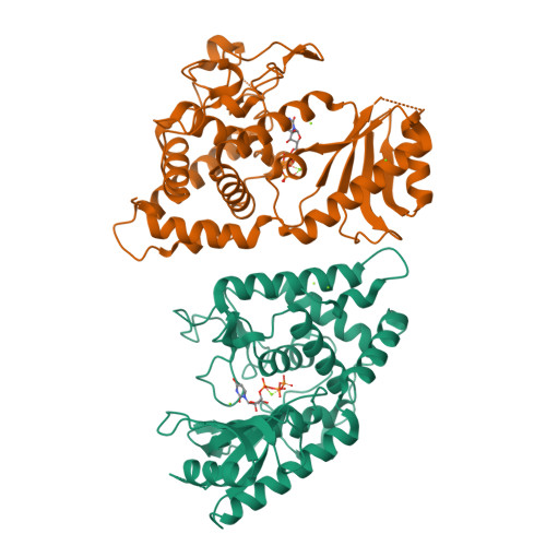
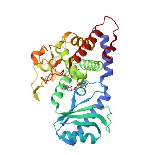
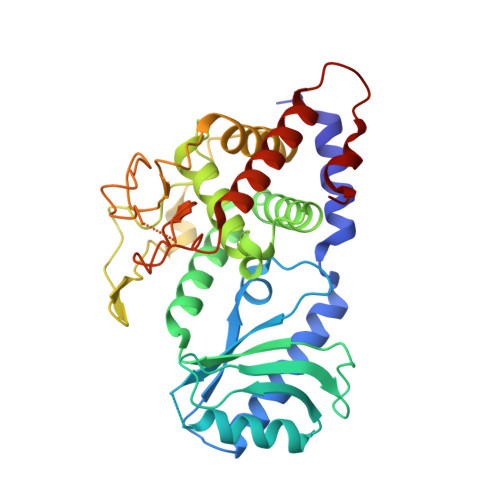
    In eukaryotes, mRNA degradation begins with poly(A) tail removal, followed by decapping, and the mRNA body is degraded by exonucleases. In recent years, the major influence of 3'-end uridylation as a regulatory step within several RNA degradation pathways has generated significant attention toward the responsible enzymes, which are called poly(U) polymerases (PUPs). We determined the atomic structure of the Cid1 protein, the founding member of the PUP family, in its UTP-bound form, allowing unambiguous positioning of the UTP molecule. Our data also suggest that the RNA substrate accommodation and product translocation by the Cid1 protein rely on local and global movements of the enzyme. Supplemented by point mutations, the atomic model is used to propose a catalytic cycle. Our study underlines the Cid1 RNA binding properties, a feature with critical implications for miRNAs, histone mRNAs, and, more generally, cellular RNA degradation.


  • Organizational Affiliation

    Department of Molecular Biology, University of Geneva, 30 Quai Ernest Ansermet, Geneva 1211, Switzerland.


Macromolecules
Find similar proteins by:  (by identity cutoff)  |  3D Structure
Entity ID: 1
MoleculeChains Sequence LengthOrganismDetailsImage
Poly(A) RNA polymerase protein cid1
A, B
340Schizosaccharomyces pombe 972h-Mutation(s): 0 
Gene Names: cid1SPAC19D5.03
EC: 2.7.7 (PDB Primary Data), 2.7.7.19 (UniProt), 2.7.7.52 (UniProt)
UniProt
Find proteins for O13833 (Schizosaccharomyces pombe (strain 972 / ATCC 24843))
Explore O13833 
Go to UniProtKB:  O13833
Entity Groups  
Sequence Clusters30% Identity50% Identity70% Identity90% Identity95% Identity100% Identity
UniProt GroupO13833
Sequence Annotations
Expand
  • Reference Sequence
Small Molecules
Ligands 2 Unique
IDChains Name / Formula / InChI Key2D Diagram3D Interactions
UTP
Query on UTP

Download Ideal Coordinates CCD File 
C [auth A],
H [auth B]
URIDINE 5'-TRIPHOSPHATE
C9 H15 N2 O15 P3
PGAVKCOVUIYSFO-XVFCMESISA-N
MG
Query on MG

Download Ideal Coordinates CCD File 
D [auth A]
E [auth A]
F [auth A]
G [auth A]
I [auth B]
MAGNESIUM ION
Mg
JLVVSXFLKOJNIY-UHFFFAOYSA-N
Experimental Data & Validation

Experimental Data

  • Method: X-RAY DIFFRACTION
  • Resolution: 2.28 Å
  • R-Value Free:  0.247 (Depositor), 0.240 (DCC) 
  • R-Value Work:  0.188 (Depositor), 0.180 (DCC) 
  • R-Value Observed: 0.191 (Depositor) 
Space Group: P 1 21 1
Unit Cell:
Length ( Å )Angle ( ˚ )
a = 53.49α = 90
b = 76.86β = 91.17
c = 81.86γ = 90
Software Package:
Software NamePurpose
ADSCdata collection
PHASERphasing
PHENIXrefinement
XDSdata reduction
XDSdata scaling

Structure Validation

View Full Validation Report


Ligand Structure Quality Assessment 

Created with Raphaël 2.3.0Worse 01 BetterLigand structure goodness of fit to experimental dataBest fitted UTPClick on this verticalbar to view details

Entry History 

Deposition Data

Revision History  (Full details and data files)

  • Version 1.0: 2012-06-06
    Type: Initial release
  • Version 1.1: 2012-06-27
    Changes: Database references
  • Version 1.2: 2024-02-28
    Changes: Data collection, Database references, Derived calculations