4AKB

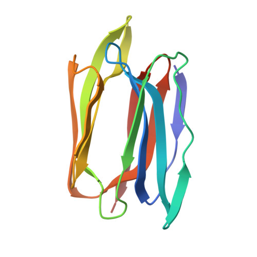
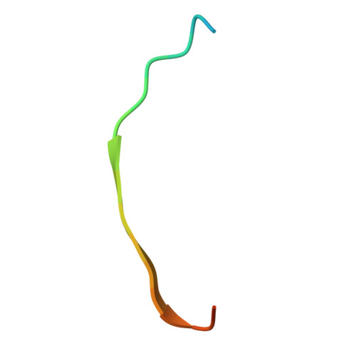
Structure of Galactose Binding lectin from Champedak (CGB) with Galactose


Experimental Data Snapshot

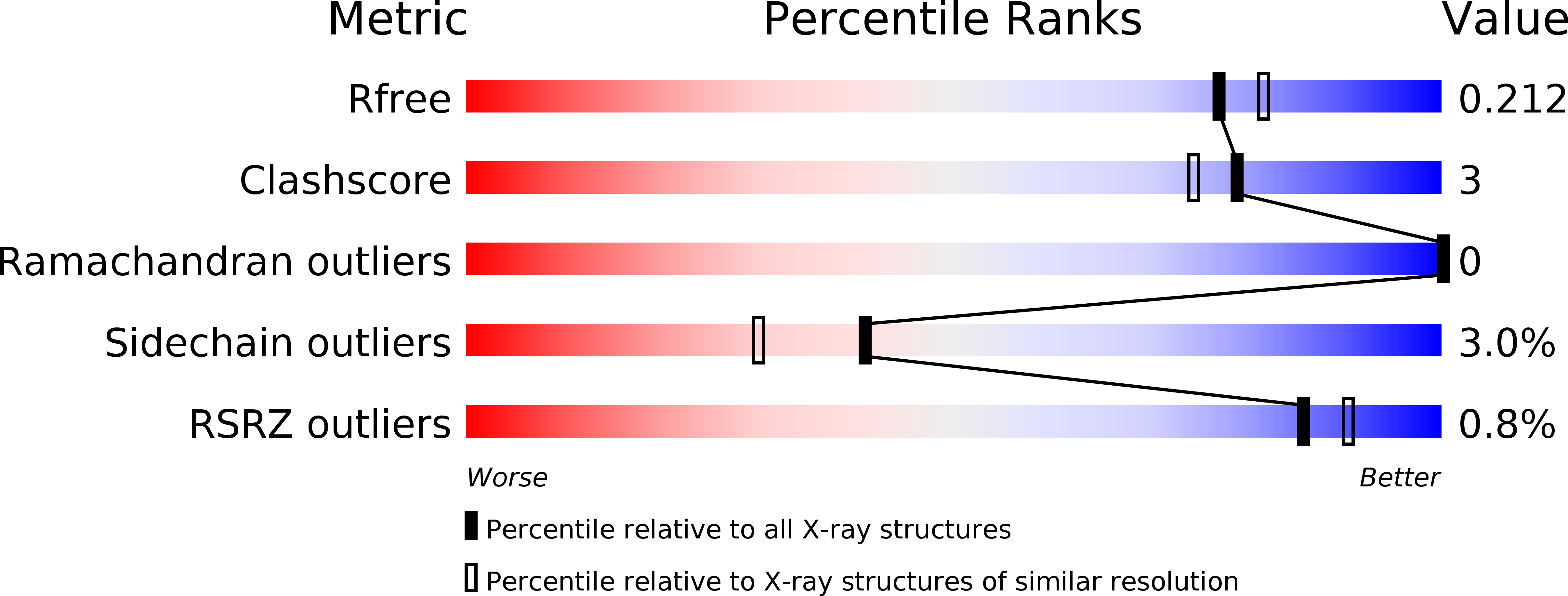
  • Method: X-RAY DIFFRACTION
  • Resolution: 1.95 Å
  • R-Value Free: 0.197 
  • R-Value Work: 0.171 
  • R-Value Observed: 0.172 

Starting Model: experimental
View more details

wwPDB Validation   3D Report Full Report


Ligand Structure Quality Assessment 


This is version 1.4 of the entry. See complete history


Literature

Structures and Binding Specificity of Galactose- and Mannose-Binding Lectins from Champedak: Differences from Jackfruit Lectins

Gabrielsen, M.Abdul-Rahman, P.S.Othman, S.Hashim, O.H.Cogdell, R.J.

(2014) Acta Crystallogr Sect F Struct Biol Cryst Commun 70: 709

  • DOI: https://doi.org/10.1107/S2053230X14008966
  • Primary Citation of Related Structures:  
    4AK4, 4AKB, 4AKC, 4AKD

  • PubMed Abstract: 

    Galactose-binding and mannose-binding lectins from the champedak fruit, which is native to South-east Asia, exhibit useful potential clinical applications. The specificity of the two lectins for their respective ligands allows the detection of potential cancer biomarkers and monitoring of the glycosylated state of proteins in human serum and/or urine. To fully understand and expand the use of these natural proteins, their complete sequences and crystal structures are presented here, together with details of sugar binding.


  • Organizational Affiliation

    Institute of Molecular, Cell and Systems Biology, University of Glasgow, 120 University Avenue, Glasgow G12 8TA, Scotland.


Macromolecules
Find similar proteins by:  (by identity cutoff)  |  3D Structure
Entity ID: 1
MoleculeChains Sequence LengthOrganismDetailsImage
AGGLUTININ ALPHA CHAIN
A, C, E, G
133Artocarpus integerMutation(s): 0 
UniProt
Find proteins for P18670 (Artocarpus integer)
Explore P18670 
Go to UniProtKB:  P18670
Entity Groups  
Sequence Clusters30% Identity50% Identity70% Identity90% Identity95% Identity100% Identity
UniProt GroupP18670
Sequence Annotations
Expand
  • Reference Sequence

Find similar proteins by:  Sequence   |   3D Structure  

Entity ID: 2
MoleculeChains Sequence LengthOrganismDetailsImage
AGGLUTININ BETA-4 CHAIN
B, D, F, H
21Artocarpus integerMutation(s): 0 
UniProt
Find proteins for Q9S8T0 (Artocarpus integer)
Explore Q9S8T0 
Go to UniProtKB:  Q9S8T0
Entity Groups  
Sequence Clusters30% Identity50% Identity70% Identity90% Identity95% Identity100% Identity
UniProt GroupQ9S8T0
Sequence Annotations
Expand
  • Reference Sequence
Experimental Data & Validation

Experimental Data

  • Method: X-RAY DIFFRACTION
  • Resolution: 1.95 Å
  • R-Value Free: 0.197 
  • R-Value Work: 0.171 
  • R-Value Observed: 0.172 
  • Space Group: P 21 21 2
Unit Cell:
Length ( Å )Angle ( ˚ )
a = 76.286α = 90
b = 121.03β = 90
c = 77.638γ = 90
Software Package:
Software NamePurpose
BUSTERrefinement
MOSFLMdata reduction
SCALAdata scaling
PHASERphasing

Structure Validation

View Full Validation Report


Ligand Structure Quality Assessment 


Entry History 

Deposition Data

Revision History  (Full details and data files)

  • Version 1.0: 2013-02-27
    Type: Initial release
  • Version 1.1: 2014-06-11
    Changes: Database references
  • Version 1.2: 2014-06-18
    Changes: Database references
  • Version 1.3: 2020-07-29
    Type: Remediation
    Reason: Carbohydrate remediation
    Changes: Advisory, Data collection, Derived calculations, Other, Structure summary
  • Version 1.4: 2023-12-20
    Changes: Advisory, Data collection, Database references, Refinement description, Structure summary