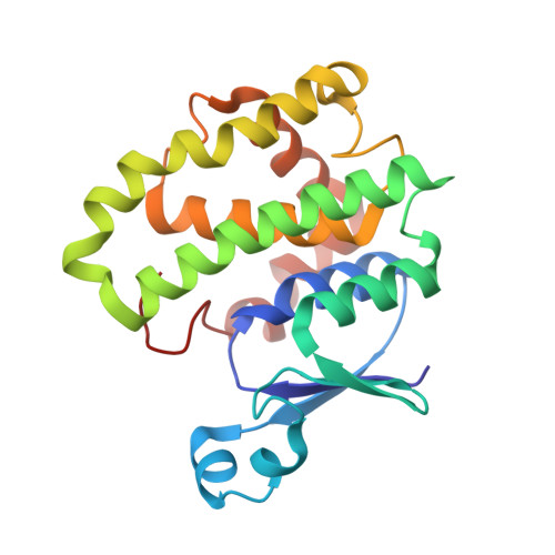
Site-directed mutagenesis of mouse glutathione transferase P1-1 unlocks masked cooperativity, introduces a novel mechanism for 'ping pong' kinetic behaviour, and provides further structural evidence for participation of a water molecule in proton abstraction from glutathione.
McManus, G., Costa, M., Canals, A., Coll, M., Mantle, T.J.(2011) FEBS J 278: 273-281
- PubMed: 21134126
- DOI: https://doi.org/10.1111/j.1742-4658.2010.07944.x
- Primary Citation of Related Structures:
3O76 - PubMed Abstract:
Mouse liver glutathione transferase P1-1 has three cysteine residues at positions 14, 47 and 169. We have constructed the single, double and triple cysteine to alanine mutants to define the behaviour of all three thiols. We confirm that C47 is the 'fast' thiol (pK 7.4), and define C169 as the alkaline reactive residue with a pK(a) of 8.6. Only a small proportion of C14 is reactive with 5,5'-dithiobis-(2-nitrobenoic acid) (DTNB) at pH 9 in the C47A/C169A double mutant. The native enzyme and the C169A mutant exhibited Michaelis-Menten kinetics, but all other thiol to alanine mutants exhibited sigmoidal kinetics to varying degrees. The C169A mutant exhibited 'ping pong' kinetics, consistent with a mechanism whereby liberation of a proton from a reduced enzyme-glutathione (GSH) complex to form an enzyme-GS(-) (unprotonated) complex is essentially irreversible. Intriguingly, similar behaviour has recently been reported for a mutant of the yeast prion Ure2p. This cooperative behaviour is 'mirrored' in the crystal structure of the C47A mutant, which binds the p-nitrobenzyl moiety of p-nitrobenzyglutathione in distinct orientations in the two crystallographic subunits. The asymmetry seen in this structure for product binding is associated with absence of a water molecule W0 in the standard wild-type conformation of product binding that is clearly identifiable in the new structure, which may represent a structural model for binding of incoming GSH prior to displacement of W0. Elimination of W0 as a hydroxonium ion may be the mechanism for the initial proton extrusion from the active site.
Organizational Affiliation:
School of Biochemistry and Immunology, Trinity College, Dublin, Ireland. [email protected]


