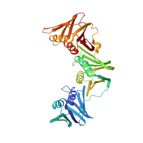
3BEP

Structure of a sliding clamp on DNA


Experimental Data Snapshot

  • Method: X-RAY DIFFRACTION
  • Resolution: 1.92 Å
  • R-Value Free: 0.268 
  • R-Value Work: 0.210 

Starting Model: experimental
View more details

wwPDB Validation   3D Report Full Report


Ligand Structure Quality Assessment 


This is version 1.3 of the entry. See complete history


Literature

Structure of a sliding clamp on DNA

Georgescu, R.E.Kim, S.S.Yurieva, O.Kuriyan, J.Kong, X.-P.O'Donnell, M.

(2008) Cell 132: 43-54

  • DOI: https://doi.org/10.1016/j.cell.2007.11.045
  • Primary Citation of Related Structures:  
    3BEP

  • PubMed Abstract: 

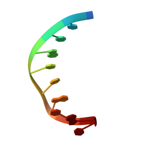
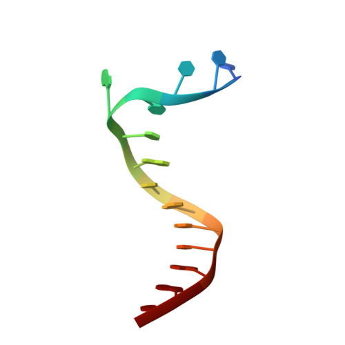
    The structure of the E. coli beta clamp polymerase processivity factor has been solved in complex with primed DNA. Interestingly, the clamp directly binds the DNA duplex and also forms a crystal contact with the ssDNA template strand, which binds into the protein-binding pocket of the clamp. We demonstrate that these clamp-DNA interactions function in clamp loading, perhaps by inducing the ring to close around DNA. Clamp binding to template ssDNA may also serve to hold the clamp at a primed site after loading or during switching of multiple factors on the clamp. Remarkably, the DNA is highly tilted as it passes through the beta ring. The pronounced 22 degrees angle of DNA through beta may enable DNA to switch between multiple factors bound to a single clamp simply by alternating from one protomer of the ring to the other.


  • Organizational Affiliation

    Howard Hughes Medical Institute, Rockefeller University, 1230 York Avenue, Box 228, New York, NY 10021, USA.


Macromolecules

Find similar proteins by:  (by identity cutoff)  |  3D Structure
Entity ID: 3
MoleculeChains Sequence LengthOrganismDetailsImage
DNA polymerase III subunit betaC [auth A],
D [auth B]
366Escherichia coliMutation(s): 0 
Gene Names: dnaN
EC: 2.7.7.7
UniProt
Find proteins for P0A988 (Escherichia coli (strain K12))
Explore P0A988 
Go to UniProtKB:  P0A988
Entity Groups  
Sequence Clusters30% Identity50% Identity70% Identity90% Identity95% Identity100% Identity
UniProt GroupP0A988
Sequence Annotations
Expand
  • Reference Sequence

Find similar nucleic acids by:  Sequence   |   3D Structure  

Entity ID: 1
MoleculeChains LengthOrganismImage
DNA (5'-D(P*DCP*DCP*DCP*DAP*DTP*DCP*DGP*DTP*DAP*DT)-3')A [auth C]10N/A
Sequence Annotations
Expand
  • Reference Sequence

Find similar nucleic acids by:  Sequence   |   3D Structure  

Entity ID: 2
MoleculeChains LengthOrganismImage
DNA (5'-D(*DTP*DTP*DTP*DTP*DAP*DTP*DAP*DCP*DGP*DAP*DTP*DGP*DGP*DG)-3')B [auth D]14N/A
Sequence Annotations
Expand
  • Reference Sequence
Small Molecules
Ligands 1 Unique
IDChains Name / Formula / InChI Key2D Diagram3D Interactions
5CY
Query on 5CY

Download Ideal Coordinates CCD File 
E [auth C]1-(3-hydroxypropyl)-2-{(1E,3E,5E)-5-[1-(3-hydroxypropyl)-3,3-dimethyl-1,3-dihydro-2H-indol-2-ylidene]penta-1,3-dien-1-y l}-3,3-dimethyl-3H-indolium
C31 H39 N2 O2
VFBDPICHYNKCQJ-UHFFFAOYSA-N
Experimental Data & Validation

Experimental Data

  • Method: X-RAY DIFFRACTION
  • Resolution: 1.92 Å
  • R-Value Free: 0.268 
  • R-Value Work: 0.210 
  • Space Group: P 1
Unit Cell:
Length ( Å )Angle ( ˚ )
a = 40.203α = 113.284
b = 70.08β = 92.073
c = 73.872γ = 99.361
Software Package:
Software NamePurpose
DENZOdata reduction
SCALEPACKdata scaling
CNSrefinement
PDB_EXTRACTdata extraction
ADSCdata collection
CNSphasing

Structure Validation

View Full Validation Report


Ligand Structure Quality Assessment 


Entry History 

Deposition Data

Revision History  (Full details and data files)

  • Version 1.0: 2008-01-29
    Type: Initial release
  • Version 1.1: 2011-07-13
    Changes: Version format compliance
  • Version 1.2: 2017-10-25
    Changes: Advisory, Refinement description
  • Version 1.3: 2023-08-30
    Changes: Data collection, Database references, Derived calculations, Refinement description, Structure summary