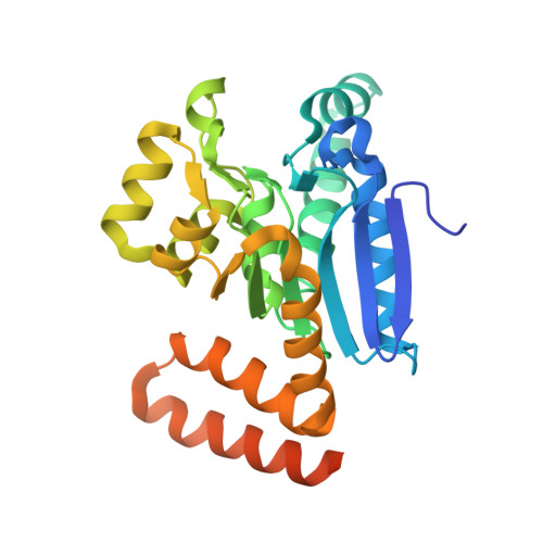
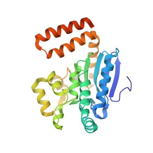
2J5I

Crystal Structure of Hydroxycinnamoyl-CoA Hydratase-Lyase


Experimental Data Snapshot

  • Method: X-RAY DIFFRACTION
  • Resolution: 1.80 Å
  • R-Value Free: 0.215 
  • R-Value Work: 0.179 
  • R-Value Observed: 0.181 

Starting Model: experimental
View more details

wwPDB Validation   3D Report Full Report


This is version 1.3 of the entry. See complete history


Literature

The 1.8 A Resolution Structure of Hydroxycinnamoyl- Coenzyme a Hydratase-Lyase (Hchl) from Pseudomonas Fluorescens, an Enzyme that Catalyses the Transformation of Feruloyl-Coenzyme a to Vanillin.

Leonard, P.M.Brzozowski, A.M.Lebedev, A.Marshall, C.M.Smith, D.J.Verma, C.S.Walton, N.J.Grogan, G.

(2006) Acta Crystallogr D Biol Crystallogr 62: 1494

  • DOI: https://doi.org/10.1107/S0907444906039199
  • Primary Citation of Related Structures:  
    2J5I

  • PubMed Abstract: 

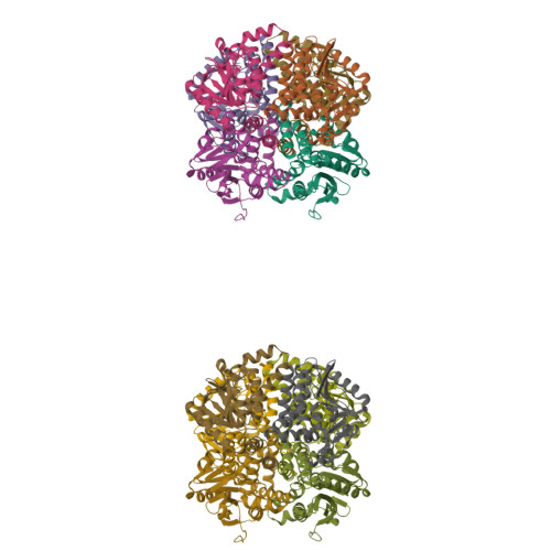
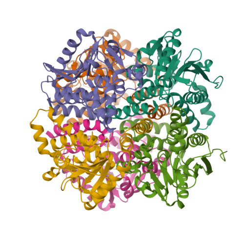
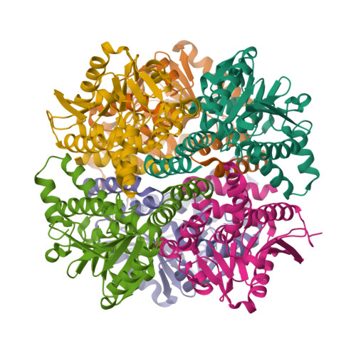
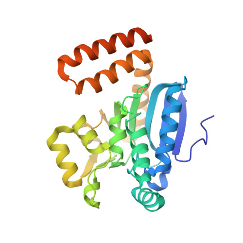
    The crystal structure of hydroxycinnamoyl-CoA hydratase-lyase (HCHL) from Pseudomonas fluorescens AN103 has been solved to 1.8 A resolution. HCHL is a member of the crotonase superfamily and catalyses the hydration of the acyl-CoA thioester of ferulic acid [3-(4-hydroxy-3-methoxy-phenyl)prop-2-enoic acid] and the subsequent retro-aldol cleavage of the hydrated intermediate to yield vanillin (4-hydroxy-3-methoxy-benzaldehyde). The structure contains 12 molecules in the asymmetric unit, in which HCHL assumes a hexameric structure of two stacked trimers. The substrate, feruloyl-CoA, was modelled into the active site based on the structure of enoyl-CoA hydratase bound to the feruloyl-CoA-like substrate 4-(N,N-dimethylamino)-cinnamoyl-CoA (PDB code 1ey3). Feruloyl-CoA was bound in this model between helix 3 of the A subunit and helix 9 of the B subunit. A highly ordered structural water in the HCHL structure coincided with the thioester carbonyl of feruloyl-CoA in the model, suggesting that the oxyanion hole for stabilization of a thioester-derived enolate, characteristic of coenzyme-A dependent members of the crotonase superfamily, is conserved. The model also suggested that a strong hydrogen bond between the phenolic hydroxyl groups of feruloyl-CoA and BTyr239 may be an important determinant of the enzyme's ability to discriminate between the natural substrate and cinnamoyl-CoA, which is not a substrate.


  • Organizational Affiliation

    Structural Biology Laboratory, Department of Chemistry, University of York, Heslington, York YO10 5YW, England.


Macromolecules
Find similar proteins by:  (by identity cutoff)  |  3D Structure
Entity ID: 1
MoleculeChains Sequence LengthOrganismDetailsImage
P-HYDROXYCINNAMOYL COA HYDRATASE/LYASE276Pseudomonas fluorescensMutation(s): 0 
EC: 4.2.1.101 (PDB Primary Data), 4.1.2.61 (UniProt)
UniProt
Find proteins for O69762 (Pseudomonas fluorescens)
Explore O69762 
Go to UniProtKB:  O69762
Entity Groups  
Sequence Clusters30% Identity50% Identity70% Identity90% Identity95% Identity100% Identity
UniProt GroupO69762
Sequence Annotations
Expand
  • Reference Sequence
Find similar proteins by:  (by identity cutoff)  |  3D Structure
Entity ID: 2
MoleculeChains Sequence LengthOrganismDetailsImage
P-HYDROXYCINNAMOYL COA HYDRATASE/LYASE
B, C, D, E, F
B, C, D, E, F, G, H, J, K, L
276Pseudomonas fluorescensMutation(s): 0 
EC: 4.2.1.101 (PDB Primary Data), 4.1.2.61 (UniProt)
UniProt
Find proteins for O69762 (Pseudomonas fluorescens)
Explore O69762 
Go to UniProtKB:  O69762
Entity Groups  
Sequence Clusters30% Identity50% Identity70% Identity90% Identity95% Identity100% Identity
UniProt GroupO69762
Sequence Annotations
Expand
  • Reference Sequence
Find similar proteins by:  (by identity cutoff)  |  3D Structure
Entity ID: 3
MoleculeChains Sequence LengthOrganismDetailsImage
P-HYDROXYCINNAMOYL COA HYDRATASE/LYASE276Pseudomonas fluorescensMutation(s): 0 
EC: 4.2.1.101 (PDB Primary Data), 4.1.2.61 (UniProt)
UniProt
Find proteins for O69762 (Pseudomonas fluorescens)
Explore O69762 
Go to UniProtKB:  O69762
Entity Groups  
Sequence Clusters30% Identity50% Identity70% Identity90% Identity95% Identity100% Identity
UniProt GroupO69762
Sequence Annotations
Expand
  • Reference Sequence
Experimental Data & Validation

Experimental Data

  • Method: X-RAY DIFFRACTION
  • Resolution: 1.80 Å
  • R-Value Free: 0.215 
  • R-Value Work: 0.179 
  • R-Value Observed: 0.181 
  • Space Group: P 21 21 2
Unit Cell:
Length ( Å )Angle ( ˚ )
a = 154.238α = 90
b = 167.487β = 90
c = 130.818γ = 90
Software Package:
Software NamePurpose
REFMACrefinement
DENZOdata reduction
SCALEPACKdata scaling
MOLREPphasing

Structure Validation

View Full Validation Report


Entry History 

Deposition Data

Revision History  (Full details and data files)

  • Version 1.0: 2006-12-06
    Type: Initial release
  • Version 1.1: 2011-05-08
    Changes: Version format compliance
  • Version 1.2: 2011-07-13
    Changes: Version format compliance
  • Version 1.3: 2023-12-13
    Changes: Data collection, Database references, Other, Refinement description