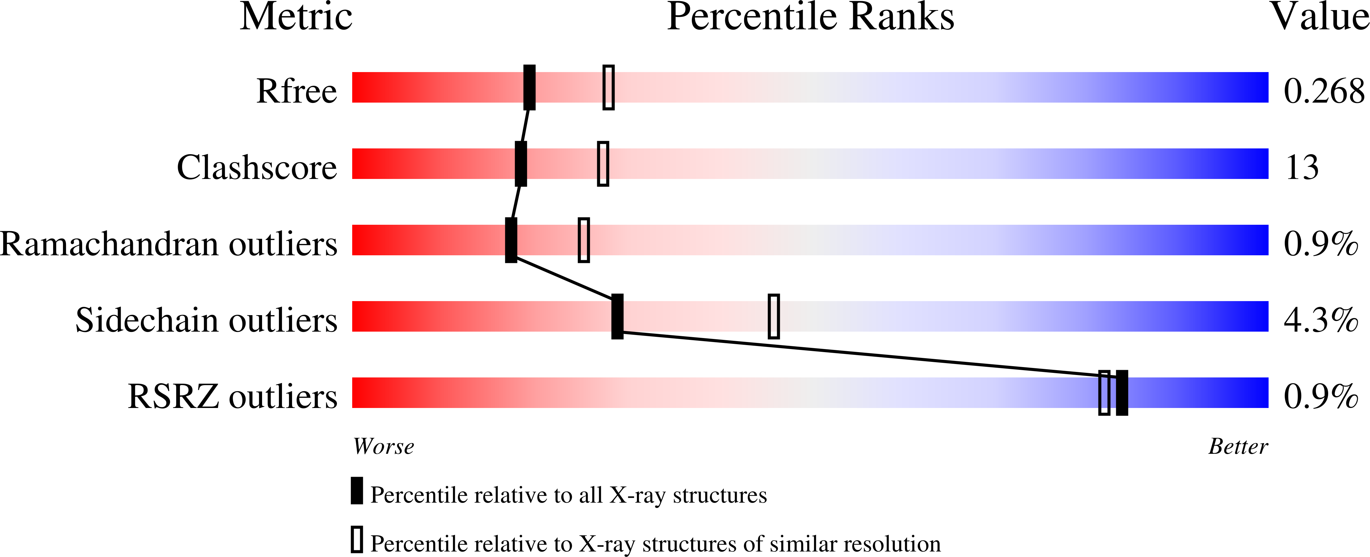
Probing the Structural Basis for the Difference in Thermostability Displayed by Family 10 Xylanases.
Xie, H., Flint, J., Vardakou, M., Lakey, J.H., Lewis, R.J., Gilbert, H.J., Dumon, C.(2006) J Mol Biol 360: 157
- PubMed: 16762367
- DOI: https://doi.org/10.1016/j.jmb.2006.05.002
- Primary Citation of Related Structures:
2CNC - PubMed Abstract:
Thermostability is an important property of industrially significant hydrolytic enzymes: understanding the structural basis for this attribute will underpin the future biotechnological exploitation of these biocatalysts. The Cellvibrio family 10 (GH10) xylanases display considerable sequence identity but exhibit significant differences in thermostability; thus, these enzymes represent excellent models to examine the structural basis for the variation in stability displayed by these glycoside hydrolases. Here, we have subjected the intracellular Cellvibrio mixtus xylanase CmXyn10B to forced protein evolution. Error-prone PCR and selection identified a double mutant, A334V/G348D, which confers an increase in thermostability. The mutant has a Tm 8 degrees C higher than the wild-type enzyme and, at 55 degrees C, the first-order rate constant for thermal inactivation of A334V/G348D is 4.1 x 10(-4) min(-1), compared to a value of 1.6 x 10(-1) min(-1) for the wild-type enzyme. The introduction of the N to C-terminal disulphide bridge into A334V/G348D, which increases the thermostability of wild-type CmXyn10B, conferred a further approximately 2 degrees C increase in the Tm of the double mutant. The crystal structure of A334V/G348D showed that the introduction of Val334 fills a cavity within the hydrophobic core of the xylanase, increasing the number of van der Waals interactions with the surrounding aromatic residues, while O(delta1) of Asp348 makes an additional hydrogen bond with the amide of Gly344 and O(delta2) interacts with the arabinofuranose side-chain of the xylose moiety at the -2 subsite. To investigate the importance of xylan decorations in productive substrate binding, the activity of wild-type CmXyn10B, the mutant A334V/G348D, and several other GH10 xylanases against xylotriose and xylotriose containing an arabinofuranose side-chain (AX3) was assessed. The enzymes were more active against AX3 than xylotriose, providing evidence that the arabinose side-chain makes a generic contribution to substrate recognition by GH10 xylanases.
Organizational Affiliation:
The Department of Animal Science, Rongchang Campus, Southwest University, The People's Republic of China, 402460.


