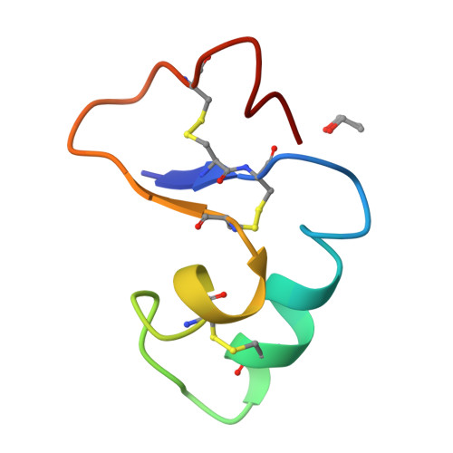
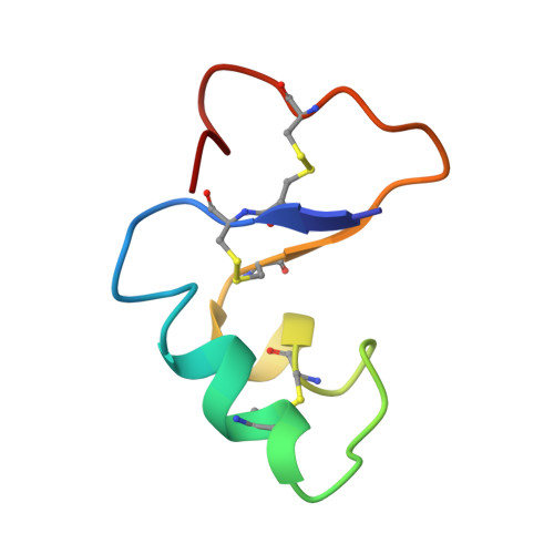
Crystal structure of Ser-22/Ile-25 form crambin confirms solvent, side chain substate correlations.
Yamano, A., Heo, N.H., Teeter, M.M.(1997) J Biological Chem 272: 9597-9600
- PubMed: 9092482
- DOI: https://doi.org/10.1074/jbc.272.15.9597
- Primary Citation of Related Structures:
1AB1 - PubMed Abstract:
It is not agreed that correlated positions of disordered protein side chains (substate correlations) can be deduced from diffraction data. The pure Ser-22/Ile-25 (SI form) crambin crystal structure confirms correlations deduced for the natural, mixed sequence form of crambin crystals. Physical separation of the mixed form into pure SI form and Pro-22/Leu-25 (PL form) crambin and the PL form crystal structure determination (Yamano, A., and Teeter, M. M. (1994) J. Biol. Chem. 269, 13956-13965) support the proposed (Teeter, M. M., Roe, S. M., and Heo, N. H. (1993) J. Mol. Biol. 230, 292-311) correlation model. Electron density of mixed form crambin crystals shows four possible pairs of side chain conformations for heterogeneous residue 22 and nearby Tyr-29 (2(2) = 4, two conformations for each of two side chains). One combination can be eliminated because of short van der Waals' contacts. However, only two alternates have been postulated to exist in mixed form crambin: Pro-22/Tyr-29A and Ser-22/Tyr-29B. In crystals of the PL form, Pro-22 and Tyr-29A are found to be in direct van der Waals' contact (Yamano, A., and Teeter, M. M. (1994) J. Biol. Chem. 269, 13956-13965). Comparison of the SI form structure with the mixed form electron density confirms that the fourth combination of side chains does not occur and that side chain correlations are mediated by water networks.
Organizational Affiliation:
X-ray Research Laboratory, Rigaku Corporation, 3-9-12 Matsubara, Akishima, 196 Tokyo, Japan.